


 癌症的基本特征包括细胞增殖、血管生成、迁移、凋亡逃避机制和细胞永生等。找到癌症发生过程中这些通路的关键标记物和对应的抗体用于检测至关重要。
 为您推荐一个泛素化位点预测神器——泛素化分析工具,可以为您的蛋白的泛素化位点作出预测和评分。
 细胞自噬受体图形绘图工具为你的蛋白的细胞受体结合位点作出预测和评分,识别结合到自噬通路中的蛋白是非常重要的,便于让我们理解自噬在正常生理、病理过程中的作用,如发育、细胞分化、神经退化性疾病、压力条件下、感染和癌症。
PCDH12 Antibody
- 产品详情
- 实验流程
- 背景知识
Application
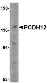
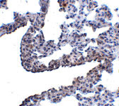
| WB, E, IHC-P |
|---|---|
| Primary Accession | Q9NPG4 |
| Other Accession | NP_057664, 7706113 |
| Reactivity | Human, Mouse, Rat |
| Host | Rabbit |
| Clonality | Polyclonal |
| Isotype | IgG |
| Calculated MW | 128995 Da |
| Concentration (mg/ml) | 1 mg/mL |
| Conjugate | Unconjugated |
| Application Notes | PCDH12 antibody can be used for detection of PCDH12 by Western blot at 2 µg/mL. Antibody can also be used for immunohistochemistry starting at 5 µg/mL. |
| Gene ID | 51294 |
|---|---|
| Other Names | Protocadherin-12, Vascular cadherin-2, Vascular endothelial cadherin-2, VE-cad-2, VE-cadherin-2, PCDH12 |
| Target/Specificity | PCDH12; This antibody is predicted to not cross-react with PCDH18. |
| Reconstitution & Storage | PCDH12 antibody can be stored at 4℃ for three months and -20℃, stable for up to one year. As with all antibodies care should be taken to avoid repeated freeze thaw cycles. Antibodies should not be exposed to prolonged high temperatures. |
| Precautions | PCDH12 Antibody is for research use only and not for use in diagnostic or therapeutic procedures. |
| Name | PCDH12 (HGNC:8657) |
|---|---|
| Function | Cellular adhesion molecule that may play an important role in cell-cell interactions at interendothelial junctions (By similarity). Acts as a regulator of cell migration, probably via increasing cell- cell adhesion (PubMed:21402705). Promotes homotypic calcium-dependent aggregation and adhesion and clusters at intercellular junctions (By similarity). Unable to bind to catenins, weakly associates with the cytoskeleton (By similarity). |
| Cellular Location | [Protocadherin-12]: Cell membrane; Single-pass type I membrane protein. Cell junction {ECO:0000250|UniProtKB:O55134} |
| Tissue Location | Expressed in highly vascularized tissues including the heart and placenta, but most tissues contain a low level of expression (PubMed:11063261). Prominent expression in the spleen (PubMed:11063261). Present in villous and extravillous trophoblast (at protein level) (PubMed:21402705). |
For Research Use Only. Not For Use In Diagnostic Procedures.
Provided below are standard protocols that you may find useful for product applications.
BACKGROUND
PCDH12 Antibody: Protocadherins comprise the largest group within the cadherin family of calcium-dependent cell-cell adhesion molecules. Protocadherin 12 (PCDH12) was initially identified through PCR screening of mouse heart microvascular endothelial cell RNA; further experiments revealed its mRNA to be strongly expressed in highly vascularized organs such as lung and kidney, in addition to glycogen-rich trophoblasts in the placenta. PCDH12-null mice are viable and fertile, but show reduced placental and embryonic sizes when compared to wild-type mice. Further studies showed significant expression changes in 2, 289 genes, including those involved in tissue morphogenesis, angiogenesis, cell-matrix adhesion and migration, immune response and chromatin remodeling.
REFERENCES
Frank M and Kemler R. Protocadherins. Curr. Opin. Cell Biol.2002; 14:557-62.
Telo P, Breviaro F, Huber P, et al. Identification of a novel cadherin (vascular endothelial cadherin-2) located at intercellular junctions in endothelial cells. J. Biol. Chem.1998; 28:17565-72.
Rampon C, Prandini MH, Bouillot S, et al. Protocadherin 12 (VE-cadherin 2) is expressed in endothelial, trophoblast, and mesangial cells. Exp. Cell Res.2005; 302:48-60.
Rampon C, Bouillot S, Climescu-Haulica A, et al. Protocadherin 12 deficiency alters morphogenesis and transcriptional profile of the placenta. Physiol. Genomics2008; 34:193-204.
终于等到您。ABCEPTA(百远生物)抗体产品。
点击下方“我要评价 ”按钮提交您的反馈信息,您的反馈和评价是我们最宝贵的财富之一,
我们将在1-3个工作日内处理您的反馈信息。
如有疑问,联系:0512-88856768 tech-china@abcepta.com.


