


 癌症的基本特征包括细胞增殖、血管生成、迁移、凋亡逃避机制和细胞永生等。找到癌症发生过程中这些通路的关键标记物和对应的抗体用于检测至关重要。
 为您推荐一个泛素化位点预测神器——泛素化分析工具,可以为您的蛋白的泛素化位点作出预测和评分。
 细胞自噬受体图形绘图工具为你的蛋白的细胞受体结合位点作出预测和评分,识别结合到自噬通路中的蛋白是非常重要的,便于让我们理解自噬在正常生理、病理过程中的作用,如发育、细胞分化、神经退化性疾病、压力条件下、感染和癌症。
RIPK1 Antibody
- 产品详情
- 实验流程
- 背景知识
Application
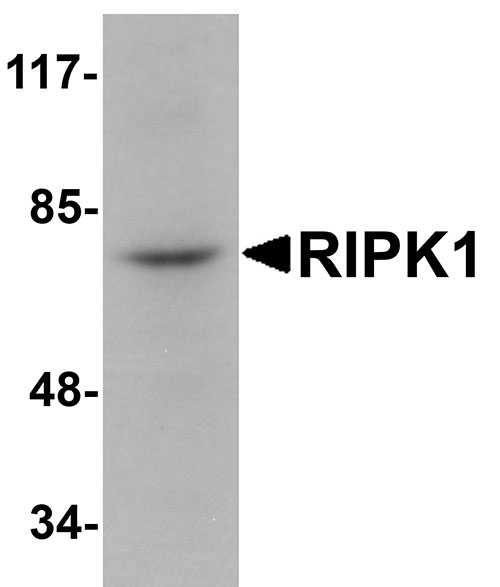
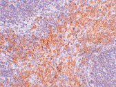
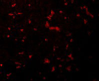
| WB, IF, E, IHC-P |
|---|---|
| Primary Accession | Q13546 |
| Other Accession | NP_003795, 57242761 |
| Reactivity | Human, Mouse, Rat |
| Host | Rabbit |
| Clonality | Polyclonal |
| Isotype | IgG |
| Calculated MW | 75931 Da |
| Concentration (mg/ml) | 1 mg/mL |
| Conjugate | Unconjugated |
| Application Notes | RIPK1 antibody can be used for detection of RIPK1 by Western blot at 1 µg/mL. Antibody can also be used for immunohistochemistry starting at 2.5 µg/mL. For immunofluorescence start at 20 µg/mL. |
| Gene ID | 8737 |
|---|---|
| Other Names | Receptor-interacting serine/threonine-protein kinase 1, 2.7.11.1, Cell death protein RIP, Receptor-interacting protein 1, RIP-1, Serine/threonine-protein kinase RIP, RIPK1, RIP, RIP1 |
| Target/Specificity | RIPK1; |
| Reconstitution & Storage | RIPK1 antibody can be stored at 4℃ for three months and -20℃, stable for up to one year. As with all antibodies care should be taken to avoid repeated freeze thaw cycles. Antibodies should not be exposed to prolonged high temperatures. |
| Precautions | RIPK1 Antibody is for research use only and not for use in diagnostic or therapeutic procedures. |
| Name | RIPK1 (HGNC:10019) |
|---|---|
| Function | Serine-threonine kinase which is a key regulator of TNF- mediated apoptosis, necroptosis and inflammatory pathways (PubMed:17703191, PubMed:24144979, PubMed:31827280, PubMed:31827281, PubMed:32657447, PubMed:35831301). Exhibits kinase activity-dependent functions that regulate cell death and kinase-independent scaffold functions regulating inflammatory signaling and cell survival (PubMed:11101870, PubMed:19524512, PubMed:19524513, PubMed:29440439, PubMed:30988283). Has kinase-independent scaffold functions: upon binding of TNF to TNFR1, RIPK1 is recruited to the TNF-R1 signaling complex (TNF-RSC also known as complex I) where it acts as a scaffold protein promoting cell survival, in part, by activating the canonical NF-kappa-B pathway (By similarity). Kinase activity is essential to regulate necroptosis and apoptosis, two parallel forms of cell death: upon activation of its protein kinase activity, regulates assembly of two death-inducing complexes, namely complex IIa (RIPK1-FADD-CASP8), which drives apoptosis, and the complex IIb (RIPK1-RIPK3-MLKL), which drives necroptosis (By similarity). RIPK1 is required to limit CASP8- dependent TNFR1-induced apoptosis (By similarity). In normal conditions, RIPK1 acts as an inhibitor of RIPK3-dependent necroptosis, a process mediated by RIPK3 component of complex IIb, which catalyzes phosphorylation of MLKL upon induction by ZBP1 (PubMed:19524512, PubMed:19524513, PubMed:29440439, PubMed:30988283). Inhibits RIPK3- mediated necroptosis via FADD-mediated recruitment of CASP8, which cleaves RIPK1 and limits TNF-induced necroptosis (PubMed:19524512, PubMed:19524513, PubMed:29440439, PubMed:30988283). Required to inhibit apoptosis and necroptosis during embryonic development: acts by preventing the interaction of TRADD with FADD thereby limiting aberrant activation of CASP8 (By similarity). In addition to apoptosis and necroptosis, also involved in inflammatory response by promoting transcriptional production of pro-inflammatory cytokines, such as interleukin-6 (IL6) (PubMed:31827280, PubMed:31827281). Phosphorylates RIPK3: RIPK1 and RIPK3 undergo reciprocal auto- and trans- phosphorylation (PubMed:19524513). Phosphorylates DAB2IP at 'Ser-728' in a TNF-alpha-dependent manner, and thereby activates the MAP3K5-JNK apoptotic cascade (PubMed:15310755, PubMed:17389591). Required for ZBP1-induced NF-kappa-B activation in response to DNA damage (By similarity). |
| Cellular Location | Cytoplasm {ECO:0000250|UniProtKB:Q60855}. Cell membrane {ECO:0000250|UniProtKB:Q9ZUF4} |
For Research Use Only. Not For Use In Diagnostic Procedures.
Provided below are standard protocols that you may find useful for product applications.
BACKGROUND
RIPK1 Antibody: RIPK1 (Receptor Interacting Protein) is a crucial 74 kD adaptor kinase in several of stress-induced signaling pathways and on the crossroad of a cell's decision to live or die. RIPK1 contains an N-terminal region with homology to protein kinases, an intermediate domain capable of association with MAPKKK and a C-terminal region containing a death domain motif present in the Fas and TNFR1 intracellular domains. Full length RIPK1 is important for signallling to NFκ-B, MAPKs and necrosis, whereas caspase-8 generates a C-terminal RIPK1 cleavage fragment, promoting TNF-induced apoptosis. It is required for TNFRSF1A-mediated and TLR3-induced NF-κB activation. RIPK1-deficient mice fail to thrive, displaying extensive apoptosis in both lymphoid and adipose tissues and dying at 1-3 days of age.
REFERENCES
Stanger BZ, Leder P, Lee TH, et al. RIP: a novel protein containing a death domain that interacts with Fas/APO-1 (CD95) in yeast and causes cell death. Cell1995; 81:513-23.
Hsu H, Huang J, Shu HB, et al. TNF-dependent recruitment of the protein kinase RIP to the TNF receptor-1 signaling complex. Immunity1996; 4:387-96.
Meylan E, Burns K, Hofmann K, et al. RIP1 is an essential mediator of Toll-like receptor 3-induced NF-kappa B activation. Nat. Immunol.2004; 5:503-7.
Festjens N, Vanden Bergh T, Cornelis S, et al. RIP1, a kinase on the crossroads of a cell's decision to live or die. Cell Death Differ.2007;14:400-10.
终于等到您。ABCEPTA(百远生物)抗体产品。
点击下方“我要评价 ”按钮提交您的反馈信息,您的反馈和评价是我们最宝贵的财富之一,
我们将在1-3个工作日内处理您的反馈信息。
如有疑问,联系:0512-88856768 tech-china@abcepta.com.


