


 癌症的基本特征包括细胞增殖、血管生成、迁移、凋亡逃避机制和细胞永生等。找到癌症发生过程中这些通路的关键标记物和对应的抗体用于检测至关重要。
 为您推荐一个泛素化位点预测神器——泛素化分析工具,可以为您的蛋白的泛素化位点作出预测和评分。
 细胞自噬受体图形绘图工具为你的蛋白的细胞受体结合位点作出预测和评分,识别结合到自噬通路中的蛋白是非常重要的,便于让我们理解自噬在正常生理、病理过程中的作用,如发育、细胞分化、神经退化性疾病、压力条件下、感染和癌症。
GOLPH2 Antibody
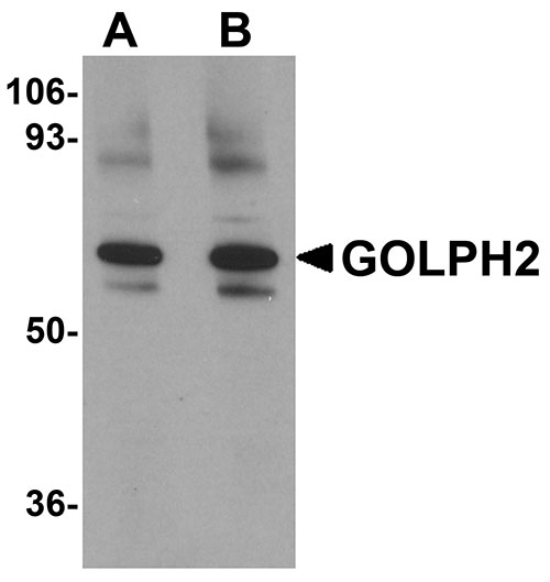
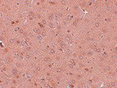
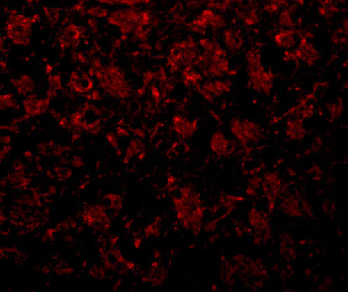
- 产品详情
- 实验流程
- 背景知识
Application 
| WB, IF, E, IHC-P |
|---|---|
| Primary Accession | Q8NBJ4 |
| Other Accession | EAW62705, 119583109 |
| Reactivity | Human, Mouse, Rat |
| Host | Rabbit |
| Clonality | Polyclonal |
| Isotype | IgG |
| Calculated MW | 45333 Da |
| Concentration (mg/ml) | 1 mg/mL |
| Conjugate | Unconjugated |
| Application Notes | GOLPH2 antibody can be used for detection of GOLPH2 by Western blot at 0.25 - 0.5 µg/mL. Antibody can also be used for immunohistochemistry starting at 2.5 µg/mL. For immunofluorescence start at 20 µg/mL. |
| Gene ID | 51280 |
|---|---|
| Other Names | Golgi membrane protein 1, Golgi membrane protein GP73, Golgi phosphoprotein 2, GOLM1, C9orf155, GOLPH2 |
| Target/Specificity | GOLM1; |
| Reconstitution & Storage | GOLPH2 antibody can be stored at 4℃ for three months and -20℃, stable for up to one year. As with all antibodies care should be taken to avoid repeated freeze thaw cycles. Antibodies should not be exposed to prolonged high temperatures. |
| Precautions | GOLPH2 Antibody is for research use only and not for use in diagnostic or therapeutic procedures. |
For Research Use Only. Not For Use In Diagnostic Procedures.
| Name | GOLM1 |
|---|---|
| Synonyms | C9orf155, GOLPH2 |
| Function | Unknown. Cellular response protein to viral infection. |
| Cellular Location | Golgi apparatus, cis-Golgi network membrane; Single-pass type II membrane protein. Note=Early Golgi. Cycles via the cell surface and endosomes upon lumenal pH disruption |
| Tissue Location | Widely expressed. Highly expressed in colon, prostate, trachea and stomach. Expressed at lower level in testis, muscle, lymphoid tissues, white blood cells and spleen. Predominantly expressed by cells of the epithelial lineage. Expressed at low level in normal liver. Expression significantly increases in virus (HBV, HCV) infected liver. Expression does not increase in liver disease due to non-viral causes (alcohol-induced liver disease, autoimmune hepatitis) Increased expression in hepatocytes appears to be a general feature of advanced liver disease. In liver tissue from patients with adult giant- cell hepatitis (GCH), it is strongly expressed in hepatocytes-derived syncytial giant cells. Constitutively expressed by biliary epithelial cells but not by hepatocytes. |
Provided below are standard protocols that you may find useful for product applications.
BACKGROUND
GOLPH2 Antibody: GOLPH2, also known as GOLM1, is a Golgi phosphoprotein that has a short cytoplasmic N-terminal domain, a membrane spanning region, and a longer C-terminal domain. It was initially identified as a possible marker for Alzheimer's disease, although later studies have demonstrated that the GOLPH2 gene does not contribute to risk of this disease. GOLPH2 expression has been reported higher in prostate cancer tissues compared to normal prostate tissue, suggesting that GOLPH2 can be used as an additional positive marker for tissue-based diagnosis of prostate cancer. It has been suggested that GOLPH2 expression in hepatocellular carcinomas (HCCs) and serum may also serve as tumor markers for HCCs.
REFERENCES
Kladney RD, Bulla GA, Guo L, et al. GP73, a novel Golgi-localized protein upregulated by viral infection. Gene 2000; 249:53-65.
Li H, Wetten S, Li L, et al. Candidate single-nucleotide polymorphisms from a genomewide association study of Alzheimer disease. Arch. Neurol. 2008; 65:45-53.
Antunez C, Boada M, Lopez-Arrieta, et al. GOLPH2 gene markers are not associated with Alzheimer’s disease in a sample of the Spanish population. J. Alzheimers Dis. 2009; 18:751-4.
Kristiansen G, Fritzsche FR, Wassermann K, et al. GOLPH2 protein expression as a novel tissue biomarker for prostate cancer: implications for tissue-based diagnostics. Br. J. Cancer 2008; 99:939-48.

终于等到您。ABCEPTA(百远生物)抗体产品。
点击下方“我要评价 ”按钮提交您的反馈信息,您的反馈和评价是我们最宝贵的财富之一,
我们将在1-3个工作日内处理您的反馈信息。
如有疑问,联系:0512-88856768 tech-china@abcepta.com.


