


 癌症的基本特征包括细胞增殖、血管生成、迁移、凋亡逃避机制和细胞永生等。找到癌症发生过程中这些通路的关键标记物和对应的抗体用于检测至关重要。
 为您推荐一个泛素化位点预测神器——泛素化分析工具,可以为您的蛋白的泛素化位点作出预测和评分。
 细胞自噬受体图形绘图工具为你的蛋白的细胞受体结合位点作出预测和评分,识别结合到自噬通路中的蛋白是非常重要的,便于让我们理解自噬在正常生理、病理过程中的作用,如发育、细胞分化、神经退化性疾病、压力条件下、感染和癌症。
RHBDD1 Antibody
- 产品详情
- 实验流程
- 背景知识
Application 
| WB, E |
|---|---|
| Primary Accession | Q8TEB9 |
| Other Accession | AAI01265, 71680537 |
| Reactivity | Human, Mouse |
| Host | Rabbit |
| Clonality | Polyclonal |
| Isotype | IgG |
| Calculated MW | 35823 Da |
| Concentration (mg/ml) | 1 mg/mL |
| Conjugate | Unconjugated |
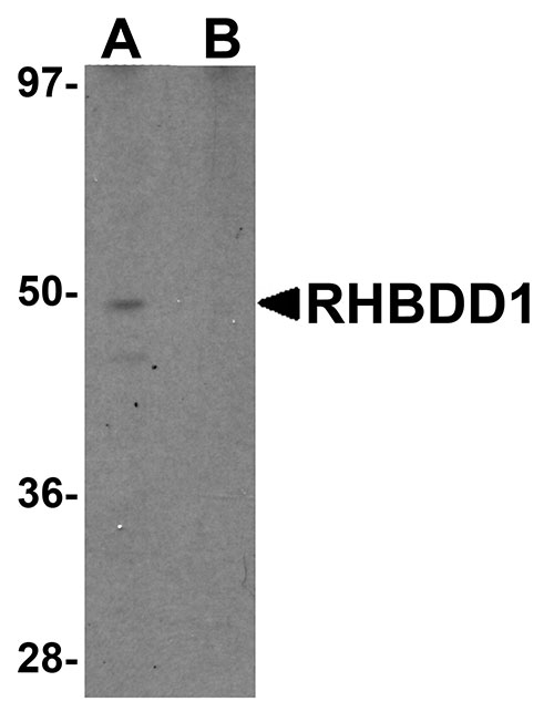
| Application Notes | RHBDD1 antibody can be used for detection of RHBDD1 by Western blot at 1 µg/mL. |
| Gene ID | 84236 |
|---|---|
| Other Names | Rhomboid-related protein 4, RRP4, 3.4.21.105, Rhomboid domain-containing protein 1, Rhomboid-like protein 4, RHBDD1, RHBDL4 |
| Target/Specificity | RHBDD1; |
| Reconstitution & Storage | RHBDD1 antibody can be stored at 4℃ for three months and -20℃, stable for up to one year. As with all antibodies care should be taken to avoid repeated freeze thaw cycles. Antibodies should not be exposed to prolonged high temperatures. |
| Precautions | RHBDD1 Antibody is for research use only and not for use in diagnostic or therapeutic procedures. |
For Research Use Only. Not For Use In Diagnostic Procedures.
| Name | RHBDD1 |
|---|---|
| Synonyms | RHBDL4 |
| Function | Intramembrane-cleaving serine protease that cleaves single transmembrane or multi-pass membrane proteins in the hydrophobic plane of the membrane, luminal loops and juxtamembrane regions. Involved in regulated intramembrane proteolysis and the subsequent release of functional polypeptides from their membrane anchors. Functional component of endoplasmic reticulum-associated degradation (ERAD) for misfolded membrane proteins. Required for the degradation process of some specific misfolded endoplasmic reticulum (ER) luminal proteins. Participates in the transfer of misfolded proteins from the ER to the cytosol, where they are destroyed by the proteasome in a ubiquitin- dependent manner. Functions in BIK, MPZ, PKD1, PTCRA, RHO, STEAP3 and TRAC processing. Involved in the regulation of exosomal secretion; inhibits the TSAP6-mediated secretion pathway. Involved in the regulation of apoptosis; modulates BIK-mediated apoptotic activity. Also plays a role in the regulation of spermatogenesis; inhibits apoptotic activity in spermatogonia. |
| Cellular Location | Endoplasmic reticulum membrane; Multi-pass membrane protein. Mitochondrion membrane; Multi-pass membrane protein |
| Tissue Location | Expressed strongly in testis. |
Provided below are standard protocols that you may find useful for product applications.
BACKGROUND
RHBDD1 Antibody: The Rhomboid family of proteins is made up of several widely conserved polytopic membrane serine proteases that play roles in growth and development. RHBDD1 is highly expressed in the testis and is involved in the cleavage of BIK, a proapoptotic member of the Bcl-2 family. Overexpression or suppression by RNAi of RHBDD1 in 293 cells will reduce or enhance BIK-mediated apoptosis, respectively, demonstrating that RHBDD1 modulates BIK-mediated apoptotic activity. In GC-1 cells, a spermatogonia cell line that can differentiate into spermatids within the seminiferous tubules, suppression of RHBDD1 expression by RNAi caused the cells to lose the ability to survive and differentiate in mouse seminiferous tubules, suggesting that RHBDD1 may be associated with mammalian spermatogenesis.
REFERENCES
Koonin EV, Makarova KS, Rogozin IB, et al. The rhomboids: a nearly ubiquitous family of intramembrane serine proteases that probably evolved by multiple horizontal gene transfers. Genome Biol.2003; 4:R19.
Wang Y, Guan X, Fok KL, et al. A novel member of the rhomboid family, RHBDD1, regulates BIK-mediated apoptosis. Cell Mol. Life Sci.2008; 65:3822-9.
Wang Y, Song W, Li S, et al. GC-1 mRHBDD1 knockdown spermatogonia cells lose their spermatogenic capacity in mouse seminiferous tubules. BNC Cell Biol.2009; 10:25.

终于等到您。ABCEPTA(百远生物)抗体产品。
点击下方“我要评价 ”按钮提交您的反馈信息,您的反馈和评价是我们最宝贵的财富之一,
我们将在1-3个工作日内处理您的反馈信息。
如有疑问,联系:0512-88856768 tech-china@abcepta.com.


