


 癌症的基本特征包括细胞增殖、血管生成、迁移、凋亡逃避机制和细胞永生等。找到癌症发生过程中这些通路的关键标记物和对应的抗体用于检测至关重要。
 为您推荐一个泛素化位点预测神器——泛素化分析工具,可以为您的蛋白的泛素化位点作出预测和评分。
 细胞自噬受体图形绘图工具为你的蛋白的细胞受体结合位点作出预测和评分,识别结合到自噬通路中的蛋白是非常重要的,便于让我们理解自噬在正常生理、病理过程中的作用,如发育、细胞分化、神经退化性疾病、压力条件下、感染和癌症。
PRDM16 Antibody

- 产品详情
- 实验流程
- 背景知识
Application
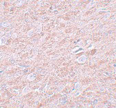
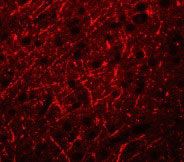
| WB, IF, E, IHC-P |
|---|---|
| Primary Accession | Q9HAZ2 |
| Other Accession | CAH71530, 55665817 |
| Reactivity | Human, Mouse, Rat |
| Host | Rabbit |
| Clonality | Polyclonal |
| Isotype | IgG |
| Calculated MW | 140251 Da |
| Concentration (mg/ml) | 1 mg/mL |
| Conjugate | Unconjugated |
| Application Notes | PRDM16 antibody can be used for detection of PRDM16 by Western blot at 1 - 2 µg/mL. Antibody can also be used for immunohistochemistry starting at 2.5 µg/mL. For immunofluorescence start at 20 µg/mL. |
| Gene ID | 63976 |
|---|---|
| Other Names | PR domain zinc finger protein 16, PR domain-containing protein 16, Transcription factor MEL1, MDS1/EVI1-like gene 1, PRDM16, KIAA1675, MEL1, PFM13 |
| Target/Specificity | PRDM16; |
| Reconstitution & Storage | PRDM16 antibody can be stored at 4℃ for three months and -20℃, stable for up to one year. As with all antibodies care should be taken to avoid repeated freeze thaw cycles. Antibodies should not be exposed to prolonged high temperatures. |
| Precautions | PRDM16 Antibody is for research use only and not for use in diagnostic or therapeutic procedures. |
| Name | PRDM16 (HGNC:14000) |
|---|---|
| Function | Binds DNA and functions as a transcriptional regulator (PubMed:12816872). Displays histone methyltransferase activity and monomethylates 'Lys-9' of histone H3 (H3K9me1) in vitro (By similarity). Probably catalyzes the monomethylation of free histone H3 in the cytoplasm which is then transported to the nucleus and incorporated into nucleosomes where SUV39H methyltransferases use it as a substrate to catalyze histone H3 'Lys-9' trimethylation (By similarity). Likely to be one of the primary histone methyltransferases along with MECOM/PRDM3 that direct cytoplasmic H3K9me1 methylation (By similarity). Functions in the differentiation of brown adipose tissue (BAT) which is specialized in dissipating chemical energy in the form of heat in response to cold or excess feeding while white adipose tissue (WAT) is specialized in the storage of excess energy and the control of systemic metabolism (By similarity). Together with CEBPB, regulates the differentiation of myoblastic precursors into brown adipose cells (By similarity). Functions as a repressor of TGF-beta signaling (PubMed:19049980). |
| Cellular Location | Nucleus. Cytoplasm |
| Tissue Location | Expressed in uterus and kidney. Expressed in both cardiomyocytes and interstitial cells. |
For Research Use Only. Not For Use In Diagnostic Procedures.
Provided below are standard protocols that you may find useful for product applications.
BACKGROUND
PRDM16 Antibody: PRDM16 is a zinc finger transcription factor and contains an N-terminal PR domain. The reciprocal translocation t(1;3)(p36;q21) occurs in a subset of myelodysplastic syndrome (MDS) and acute myeloid leukemia (AML). This gene is located near the 1p36.3 breakpoint and has been shown to be specifically expressed in the t(1:3)(p36, q21)-positive MDS/AML. The translocation results in the overexpression of a truncated version of this protein that lacks the PR domain, which may play an important role in the pathogenesis of MDS and AML. Recent studies have shown that PRDM16 normally acts as a Smad3 binding protein that may be important for the development of orofacial structures through modulation of the TGF-beta signaling pathway. Other experiments have indicated that PRDM16 controls a bidirectional cell fate switch between skeletal myoblasts and brown fat cells.
REFERENCES
Mochizuki N, Shimizu S, Nagasawa T, et al. A novel gene, MEL1, mapped to 1p36.3 is highly homologous to the MDS1/EVI1 gene and is transcriptionally activated in t(1;3)(p36;q21)-positive leukemia cells. Blood2000; 96:3209-14.
Morishita K. Leukemogenesis of the EVI1/MEL1 gene family. Int. J. Hematol.2007; 85:279-86.
Warner DR, Horn KH, Mudd L, et al. PRDM16/MEL1: a novel Smad binding protein expressed in murine embryonic orofacial tissue. Biochim. Biophys. Acta2007; 1773:814-20.
Seale P, Bjork B, Yang W, et al. PRDM16 controls a brown fat/skeletal muscle switch. Nature2008; 454:961-7.
终于等到您。ABCEPTA(百远生物)抗体产品。
点击下方“我要评价 ”按钮提交您的反馈信息,您的反馈和评价是我们最宝贵的财富之一,
我们将在1-3个工作日内处理您的反馈信息。
如有疑问,联系:0512-88856768 tech-china@abcepta.com.


