


 癌症的基本特征包括细胞增殖、血管生成、迁移、凋亡逃避机制和细胞永生等。找到癌症发生过程中这些通路的关键标记物和对应的抗体用于检测至关重要。
 为您推荐一个泛素化位点预测神器——泛素化分析工具,可以为您的蛋白的泛素化位点作出预测和评分。
 细胞自噬受体图形绘图工具为你的蛋白的细胞受体结合位点作出预测和评分,识别结合到自噬通路中的蛋白是非常重要的,便于让我们理解自噬在正常生理、病理过程中的作用,如发育、细胞分化、神经退化性疾病、压力条件下、感染和癌症。
VLK Antibody
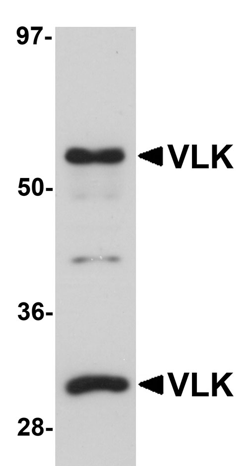
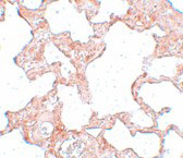
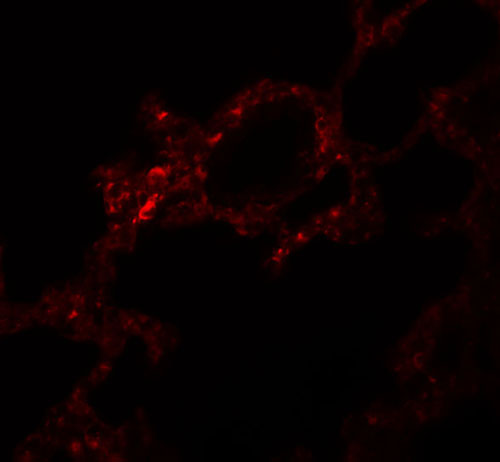
- 产品详情
- 实验流程
- 背景知识
Application 
| WB, IF, E, IHC-P |
|---|---|
| Primary Accession | Q5RJI4 |
| Other Accession | NP_612379, 238636897 |
| Reactivity | Human, Mouse, Rat |
| Host | Rabbit |
| Clonality | Polyclonal |
| Isotype | IgG |
| Calculated MW | 53841 Da |
| Concentration (mg/ml) | 1 mg/mL |
| Conjugate | Unconjugated |
| Application Notes | VLK antibody can be used for detection of VLK by Western blot at 1 µg/mL. Antibody can also be used for immunohistochemistry starting at 2.5 µg/mL. For immunofluorescence start at 5 µg/mL. |
| Gene ID | 106522 |
|---|---|
| Other Names | Extracellular tyrosine-protein kinase PKDCC, 2.7.10.2, Protein kinase domain-containing protein, cytoplasmic {ECO:0000312|MGI:MGI:2147077}, Protein kinase-like protein SgK493, Sugen kinase 493, Vertebrate lonesome kinase, Pkdcc {ECO:0000312|MGI:MGI:2147077} |
| Target/Specificity | Pkdcc; |
| Reconstitution & Storage | VLK antibody can be stored at 4℃ for three months and -20℃, stable for up to one year. As with all antibodies care should be taken to avoid repeated freeze thaw cycles. Antibodies should not be exposed to prolonged high temperatures. |
| Precautions | VLK Antibody is for research use only and not for use in diagnostic or therapeutic procedures. |
For Research Use Only. Not For Use In Diagnostic Procedures.
| Name | Pkdcc {ECO:0000312|MGI:MGI:2147077} |
|---|---|
| Function | Secreted tyrosine-protein kinase that mediates phosphorylation of extracellular proteins and endogenous proteins in the secretory pathway, which is essential for patterning at organogenesis stages. Mediates phosphorylation of MMP1, MMP13, MMP14, MMP19 and ERP29 (PubMed:25171405). May also have serine/threonine protein kinase activity (PubMed:25171405). Required for longitudinal bone growth through regulation of chondrocyte differentiation (PubMed:19097194, PubMed:23792766). May be indirectly involved in protein transport from the Golgi apparatus to the plasma membrane (PubMed:19465597). Probably plays a role in platelets: rapidly and quantitatively secreted from platelets in response to stimulation of platelet degranulation (PubMed:25171405). |
| Cellular Location | Secreted. Golgi apparatus. Note=Both secreted and present in the Golgi apparatus. |
| Tissue Location | Strongly expressed in adult heart, liver and testis with weak expression in brain, spleen, lung and thymus. In the humerus, strongly expressed in early flat proliferative chondrocytes. In the embryo, expressed in the anterior visceral endoderm and anterior primitive streak at 6.5 dpc. At 7.5 dpc, expressed in the anterior definitive endoderm (ADE) and anterior mesoderm but not in the notochord. At 8.0 dpc, expressed in the ADE and anterior embryonic mesoderm. At 8.5 dpc, expressed more broadly in anterior tissues and at the midline of the neural plate in the midbrain region as well as the lateral margins of the neural plate posterior to the metencephalic region. Also weakly expressed in the anterior mesenchyme. At 9.5 dpc, strongest expression in branchial arches and limb buds. During mid- gestation, expression continues in mesenchymal cells, particularly in areas where these cells condense. |
Provided below are standard protocols that you may find useful for product applications.
BACKGROUND
VLK Antibody: VLK was identified as a novel protein kinase that was induced after the differentiation of cultured embryonic stem cells into mesendoderm. It has no homologs in invertebrates, but is highly conserved in vertebrate species although it does not belong to any known protein kinase groups. VLK is initially expressed in E-cadherin-positive anterior visceral endoderm and mesendoderm, but its expression is later confined to E-cadherin-negative mesenchyme. It is enriched in the Golgi apparatus and is thought to regulate the rate of protein export from the Golgi. Targeted disruption of VLK in mice leads to a defect in lung development and neonatal lethality. It has been suggested that mutations in VLK may be associated with the allergic condition atopy.
REFERENCES
Kinoshita M, Era T, Jakt LM, et al. The novel protein kinase Vlk is essential for stromal function of mesenchymal cells. Development2009; 136:2069-79.
Castro-Giner F, Bustamante M, Ramon Gonzalez J, et al. A pooling-based genome-wide analysis identifies new potential candidate genes for atopy in the European Community Respiratory Health Survey (ECRHS). BMC Med. Genet.2009; 10:128.

终于等到您。ABCEPTA(百远生物)抗体产品。
点击下方“我要评价 ”按钮提交您的反馈信息,您的反馈和评价是我们最宝贵的财富之一,
我们将在1-3个工作日内处理您的反馈信息。
如有疑问,联系:0512-88856768 tech-china@abcepta.com.


