


 癌症的基本特征包括细胞增殖、血管生成、迁移、凋亡逃避机制和细胞永生等。找到癌症发生过程中这些通路的关键标记物和对应的抗体用于检测至关重要。
 为您推荐一个泛素化位点预测神器——泛素化分析工具,可以为您的蛋白的泛素化位点作出预测和评分。
 细胞自噬受体图形绘图工具为你的蛋白的细胞受体结合位点作出预测和评分,识别结合到自噬通路中的蛋白是非常重要的,便于让我们理解自噬在正常生理、病理过程中的作用,如发育、细胞分化、神经退化性疾病、压力条件下、感染和癌症。
ATP11A Antibody
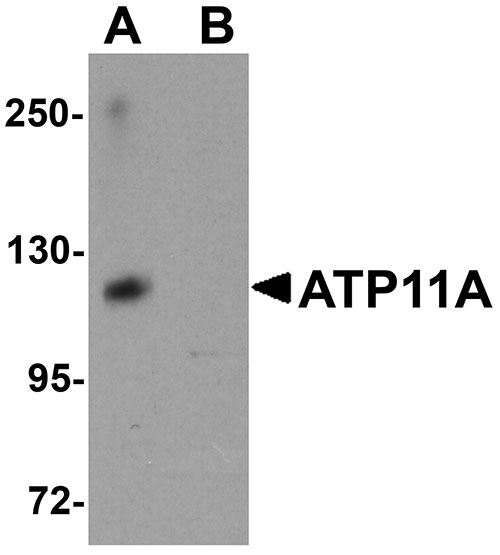

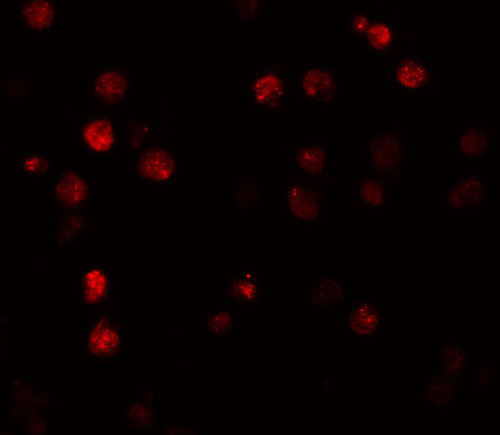
- 产品详情
- 文献引用 : 1
- 实验流程
- 背景知识
Application 
| WB, IF, ICC, E |
|---|---|
| Primary Accession | P98196 |
| Other Accession | NP_115565, 150421681 |
| Reactivity | Human, Mouse, Rat |
| Host | Rabbit |
| Clonality | Polyclonal |
| Isotype | IgG |
| Calculated MW | 129756 Da |
| Concentration (mg/ml) | 1 mg/mL |
| Conjugate | Unconjugated |
| Application Notes | ATP11A antibody can be used for detection of ATP11A by Western blot at 1 µg/mL. Antibody can also be used for immunocytochemistry starting at 10 µg/mL. For immunofluorescence start at 20 µg/mL. |
| Gene ID | 23250 |
|---|---|
| Other Names | Probable phospholipid-transporting ATPase IH, 3.6.3.1, ATPase IS, ATPase class VI type 11A, P4-ATPase flippase complex alpha subunit ATP11A, ATP11A, ATPIH, ATPIS, KIAA1021 |
| Target/Specificity | ATP11A; |
| Reconstitution & Storage | ATP11A antibody can be stored at 4℃ for three months and -20℃, stable for up to one year. As with all antibodies care should be taken to avoid repeated freeze thaw cycles. Antibodies should not be exposed to prolonged high temperatures. |
| Precautions | ATP11A Antibody is for research use only and not for use in diagnostic or therapeutic procedures. |
For Research Use Only. Not For Use In Diagnostic Procedures.
| Name | ATP11A |
|---|---|
| Synonyms | ATPIH, ATPIS, KIAA1021 |
| Function | Catalytic component of a P4-ATPase flippase complex which catalyzes the hydrolysis of ATP coupled to the transport of aminophospholipids, phosphatidylserines (PS) and phosphatidylethanolamines (PE), from the outer to the inner leaflet of the plasma membrane (PubMed:25315773, PubMed:25947375, PubMed:26567335, PubMed:29799007, PubMed:30018401, PubMed:36300302). Does not show flippase activity toward phosphatidylcholine (PC) (PubMed:34403372). Contributes to the maintenance of membrane lipid asymmetry with a specific role in morphogenesis of muscle cells. In myoblasts, mediates PS enrichment at the inner leaflet of plasma membrane, triggering PIEZO1-dependent Ca2+ influx and Rho GTPases signal transduction, subsequently leading to the assembly of cortical actomyosin fibers and myotube formation (PubMed:29799007). May be involved in the uptake of farnesyltransferase inhibitor drugs, such as lonafarnib. |
| Cellular Location | Cell membrane; Multi-pass membrane protein. Early endosome. Recycling endosome. Endoplasmic reticulum membrane; Multi-pass membrane protein. Note=Efficient exit from the endoplasmic reticulum requires the presence of CDC50A. |
| Tissue Location | Widely expressed (PubMed:26567335). Expressed in myoblasts (PubMed:29799007). |

Provided below are standard protocols that you may find useful for product applications.
BACKGROUND
ATP11A Antibody: ATP11A is a widely expressed integral membrane ATPase. ATP11A is probably phosphorylated in its intermediate state and likely drives the transport of ions such as calcium or other molecules across membranes. While the exact molecule ATP11A transports is unknown, increased expression of ATP11A mRNA has been observed in murine leukemia cells made resistant to anti-cancer drugs such as farnesyltransferase inhibitors (FTIs). Furthermore, overexpression of ATP11A provided protection against the FTI SCH66336 while knockdown of ATP11A via siRNA made cells more sensitive to this drug. Other reports suggest that elevated levels of ATP11A mRNA may also be a predictive marker of metastasis in colorectal cancer patients.
REFERENCES
Halleck MS, Lawler JF Jr, Blackshaw S, et al. Differential expression of putative transbilayer amphipath transporters. Physiol. Genomics1999; 1:139-50.
Zhang B, Groffen J, and Heisterkamp N. Resistance to farnesyltransferase inhibitors in Bcr/Abl-positive lymphoblastic leukemia by increased expression of a novel ABC transporter homolog ATP11a. Blood2005; 106:1355-61.
Miyoshi N, Ishii H, Mimori K, et al. SCRN1 is a novel marker for prognosis in colorectal cancer. J. Surg. Oncol.2010; 101:156-9.

终于等到您。ABCEPTA(百远生物)抗体产品。
点击下方“我要评价 ”按钮提交您的反馈信息,您的反馈和评价是我们最宝贵的财富之一,
我们将在1-3个工作日内处理您的反馈信息。
如有疑问,联系:0512-88856768 tech-china@abcepta.com.


