


 癌症的基本特征包括细胞增殖、血管生成、迁移、凋亡逃避机制和细胞永生等。找到癌症发生过程中这些通路的关键标记物和对应的抗体用于检测至关重要。
 为您推荐一个泛素化位点预测神器——泛素化分析工具,可以为您的蛋白的泛素化位点作出预测和评分。
 细胞自噬受体图形绘图工具为你的蛋白的细胞受体结合位点作出预测和评分,识别结合到自噬通路中的蛋白是非常重要的,便于让我们理解自噬在正常生理、病理过程中的作用,如发育、细胞分化、神经退化性疾病、压力条件下、感染和癌症。
SYNPO Antibody


- 产品详情
- 实验流程
- 背景知识
Application
| WB, IF, E, IHC-P |
|---|---|
| Primary Accession | Q8N3V7 |
| Other Accession | NP_001159680, 261278297 |
| Reactivity | Human, Mouse, Rat |
| Host | Rabbit |
| Clonality | Polyclonal |
| Isotype | IgG |
| Calculated MW | 99463 Da |
| Concentration (mg/ml) | 1 mg/mL |
| Conjugate | Unconjugated |
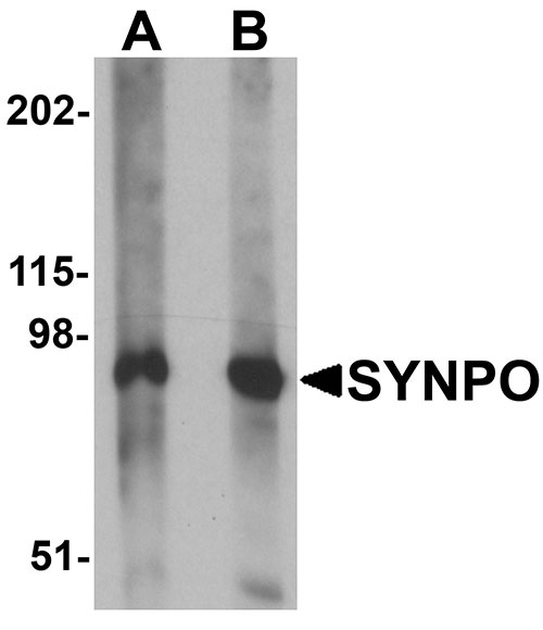
| Application Notes | SYNPO antibody can be used for detection of SYNPO by Western blot at 1 - 2 µg/mL. Antibody can also be used for immunohistochemistry starting at 2.5 µg/mL. For immunofluorescence start at 20 µg/mL. |
| Gene ID | 11346 |
|---|---|
| Other Names | Synaptopodin, SYNPO, KIAA1029 |
| Target/Specificity | SYNPO; At least three isoforms of SYNPO are known to exist; this antibody will detect all three isoforms. SYNPO antibody is predicted to not cross-react with other SYNPO family member proteins. |
| Reconstitution & Storage | SYNPO antibody can be stored at 4℃ for three months and -20℃, stable for up to one year. As with all antibodies care should be taken to avoid repeated freeze thaw cycles. Antibodies should not be exposed to prolonged high temperatures. |
| Precautions | SYNPO Antibody is for research use only and not for use in diagnostic or therapeutic procedures. |
| Name | SYNPO |
|---|---|
| Synonyms | KIAA1029 |
| Function | Actin-associated protein that may play a role in modulating actin-based shape and motility of dendritic spines and renal podocyte foot processes. Seems to be essential for the formation of spine apparatuses in spines of telencephalic neurons, which is involved in synaptic plasticity (By similarity). |
| Cellular Location | Cytoplasm, cytoskeleton {ECO:0000250|UniProtKB:Q8CC35}. Cell junction, tight junction {ECO:0000250|UniProtKB:Q8CC35}. Perikaryon {ECO:0000250|UniProtKB:Q8CC35}. Cell projection, dendritic spine {ECO:0000250|UniProtKB:Q8CC35}. Postsynaptic density {ECO:0000250|UniProtKB:Q8CC35}. Synapse {ECO:0000250|UniProtKB:Q8CC35} Cytoplasm, cytosol. Note=Localized at the tight junction of cells. In brain, localized to the postsynaptic densities and in the perikarya. Associated with dendritic spines of a subset of synapses. {ECO:0000250|UniProtKB:Q8CC35} |
| Tissue Location | Expressed in cerebral cortex. |
For Research Use Only. Not For Use In Diagnostic Procedures.
Provided below are standard protocols that you may find useful for product applications.
BACKGROUND
SYNPO Antibody: SYNPO, also known as synaptopodin, is an actin-associated protein in telencephalic dendrites and renal podocytes. SYNPO is tightly associated with the dendritic spine apparatus, and mice lacking the SYNPO gene lack the apparatus. These SYNPO-null mice also demonstrate an impaired ability to express long-term potentiation as well as deficits in spatial memory tasks, indicating that SYNPO is involved in the regulation of synaptic plasticity. Recent studies suggest that SYNPO is linked to neuronal calcium stores and plays a role in the calcium store-associated ability of neurons to undergo long-term plasticity.
REFERENCES
Mundel P, Heid HW, Mundel TM, et al. Synaptopodin: an actin-associated protein in telencephalic dendrites and renal podocytes. J. Cell Biol. 1997; 139:193-204.
Deller T, Merten T, Roth SU, et al. Actin-associated protein synaptopodin in the rat hippocampal formation: localization in the spine neck and close association with the spine apparatus of principal neurons. J. Compl. Neurol. 2000; 418:164-81.
Deller T, Korte M, Chabanis S, et al. Synaptopodin-deficient mice lack a spine apparatus and show deficits in synaptic plasticity. Proc. Natl. Acad. Sci. USA 2003; 100:10494-9.
Jedlicka P, Schwarzacher SW, Winkels R, et al. Impairment of in vivo theta-burst long-term potentiation and network excitability in the dentate gyrus of synaptopodin-deficient mice lacking the spine apparatus and the cisternal organelle. Hippocampus 2009; 19:130-40.
终于等到您。ABCEPTA(百远生物)抗体产品。
点击下方“我要评价 ”按钮提交您的反馈信息,您的反馈和评价是我们最宝贵的财富之一,
我们将在1-3个工作日内处理您的反馈信息。
如有疑问,联系:0512-88856768 tech-china@abcepta.com.


