


 癌症的基本特征包括细胞增殖、血管生成、迁移、凋亡逃避机制和细胞永生等。找到癌症发生过程中这些通路的关键标记物和对应的抗体用于检测至关重要。
 为您推荐一个泛素化位点预测神器——泛素化分析工具,可以为您的蛋白的泛素化位点作出预测和评分。
 细胞自噬受体图形绘图工具为你的蛋白的细胞受体结合位点作出预测和评分,识别结合到自噬通路中的蛋白是非常重要的,便于让我们理解自噬在正常生理、病理过程中的作用,如发育、细胞分化、神经退化性疾病、压力条件下、感染和癌症。
MYOZAP Antibody
- 产品详情
- 实验流程
- 背景知识
Application 
| WB, IF, E, IHC-P |
|---|---|
| Primary Accession | P0CAP1 |
| Other Accession | NP_001018110, 70166414 |
| Reactivity | Human, Mouse, Rat |
| Host | Rabbit |
| Clonality | Polyclonal |
| Isotype | IgG |
| Calculated MW | 54206 Da |
| Concentration (mg/ml) | 1 mg/mL |
| Conjugate | Unconjugated |
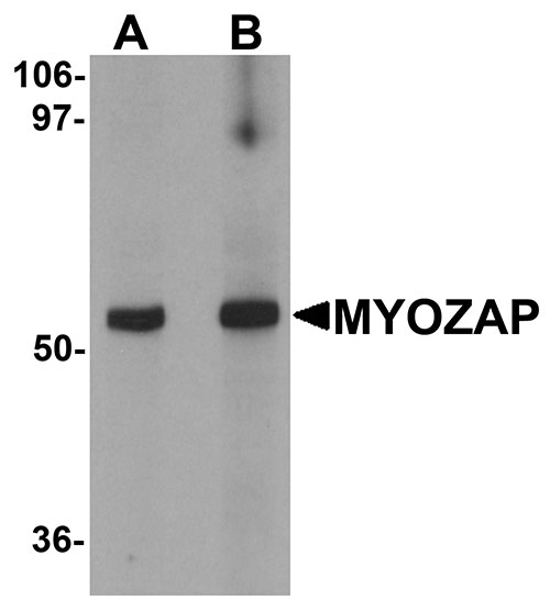
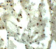
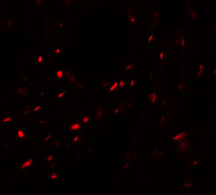
| Application Notes | MYOZAP antibody can be used for detection of MYOZAP by Western blot at 1 - 2 µg/mL. Antibody can also be used for immunohistochemistry starting at 5 µg/mL. For immunofluorescence start at 20 µg/mL. |
| Gene ID | 100820829 |
|---|---|
| Other Names | Myocardial zonula adherens protein, GRINL1A upstream protein, Gup, MYZAP, MYOZAP |
| Target/Specificity | GCOM1; Multiple isoforms of MYOZAP are known to exist. |
| Reconstitution & Storage | MYOZAP antibody can be stored at 4℃ for three months and -20℃, stable for up to one year. As with all antibodies care should be taken to avoid repeated freeze thaw cycles. Antibodies should not be exposed to prolonged high temperatures. |
| Precautions | MYOZAP Antibody is for research use only and not for use in diagnostic or therapeutic procedures. |
For Research Use Only. Not For Use In Diagnostic Procedures.
| Name | MYZAP |
|---|---|
| Synonyms | MYOZAP |
| Function | Plays a role in cellular signaling via Rho-related GTP- binding proteins and subsequent activation of transcription factor SRF (By similarity). Targets TJP1 to cell junctions. In cortical neurons, may play a role in glutaminergic signal transduction through interaction with the NMDA receptor subunit GRIN1 (By similarity). |
| Cellular Location | Cytoplasm, cytoskeleton {ECO:0000250|UniProtKB:Q5EB94}. Cell membrane {ECO:0000250|UniProtKB:Q5EB94}; Peripheral membrane protein {ECO:0000250|UniProtKB:Q5EB94}; Cytoplasmic side {ECO:0000250|UniProtKB:Q5EB94}. Cytoplasm, myofibril, sarcomere, I band {ECO:0000250|UniProtKB:Q5EB94}. Cytoplasm, myofibril, sarcomere, Z line {ECO:0000250|UniProtKB:Q5EB94}. Cell junction Note=Detected predominantly at the intercalated disk in cardiomyocytes, and at low levels on sarcomeric Z disks. Colocalizes with F-actin Colocalizes with cortical actin. {ECO:0000250|UniProtKB:Q5EB94} |
| Tissue Location | Detected in heart, liver, skeletal muscle, placenta, small intestine, lung, prostate and testis. Expressed in arrector pili muscle (at protein level) (PubMed:29034528) |
Provided below are standard protocols that you may find useful for product applications.
BACKGROUND
MYOZAP Antibody: MYOZAP, also known as GRINL1A, is a 54 kDa highly conserved cardiac protein. It is strongly expressed in the heart and lung and is a novel component of intercalated disc. MYOZAP interacts with myosin phosphatase-RhoA interacting protein (MRIP) and acts as an activator of Rho-dependent SRF signaling. Knockdown study in zebrafish results in cardiomyopathy with severe dysfunction. The MYOZAP gene is part of a complex transcript unit that includes the gene for glutamate receptor, ionotropic, N-methyl D-aspartate-like 1A (GRINL1A). Transcription of this gene occurs at an upstream promoter, with two different groups of alternatively spliced variants: Gup for GRINL1A upstream transcripts and Gcom for GRINL1A combined transcripts.
REFERENCES
Van Tintelen JP, Hofstra RM, Wiesfeld AC, et al. Molecular genetics of arrhythmogenic right ventricular cardiomyopathy: emerging horizon? Curr. Opin. Cardiol. 2007; 22:185-92.
Seeger TS, Frank D, Rohr C, et al. Myozap, a novel intercalated disc protein, activates serum response factor-dependent signaling and is required to maintain cardiac function in vivo. Circ. Res. 2010; 106:880-90.
Roginski RS, Mohan Raj BK, Birditt B, et al. The human GRINL1A gene defines a complex transcription unit, an unusual form of gene organization in eukaryotes. Genomics 2004; 84:265-76.

终于等到您。ABCEPTA(百远生物)抗体产品。
点击下方“我要评价 ”按钮提交您的反馈信息,您的反馈和评价是我们最宝贵的财富之一,
我们将在1-3个工作日内处理您的反馈信息。
如有疑问,联系:0512-88856768 tech-china@abcepta.com.


