


 癌症的基本特征包括细胞增殖、血管生成、迁移、凋亡逃避机制和细胞永生等。找到癌症发生过程中这些通路的关键标记物和对应的抗体用于检测至关重要。
 为您推荐一个泛素化位点预测神器——泛素化分析工具,可以为您的蛋白的泛素化位点作出预测和评分。
 细胞自噬受体图形绘图工具为你的蛋白的细胞受体结合位点作出预测和评分,识别结合到自噬通路中的蛋白是非常重要的,便于让我们理解自噬在正常生理、病理过程中的作用,如发育、细胞分化、神经退化性疾病、压力条件下、感染和癌症。
KCNK13 Antibody
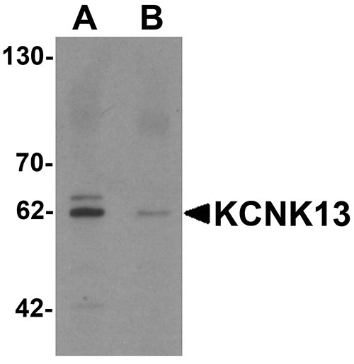
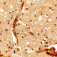
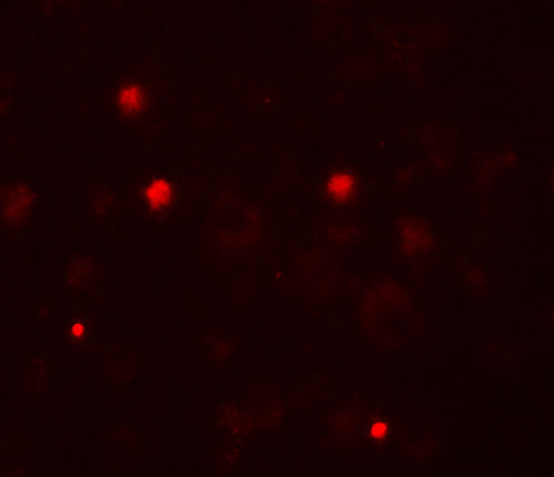
- 产品详情
- 实验流程
- 背景知识
Application 
| WB, IF, E, IHC-P |
|---|---|
| Primary Accession | Q9HB14 |
| Other Accession | NP_071337, 16306555 |
| Reactivity | Human, Mouse, Rat |
| Host | Rabbit |
| Clonality | Polyclonal |
| Isotype | IgG |
| Calculated MW | 45391 Da |
| Concentration (mg/ml) | 1 mg/mL |
| Conjugate | Unconjugated |
| Application Notes | KCNK13 antibody can be used for detection of KCNK13 by Western blot at 0.5 µg/mL. Antibody can also be used for immunohistochemistry starting at 5 µg/mL. For immunofluorescence start at 20 µg/mL. |
| Gene ID | 56659 |
|---|---|
| Other Names | Potassium channel subfamily K member 13, Tandem pore domain halothane-inhibited potassium channel 1, THIK-1, KCNK13 |
| Target/Specificity | KCNK13; KCNK13 antibody is predicted to not cross-react with other KCNK protein family members. |
| Reconstitution & Storage | KCNK13 antibody can be stored at 4℃ for three months and -20℃, stable for up to one year. As with all antibodies care should be taken to avoid repeated freeze thaw cycles. Antibodies should not be exposed to prolonged high temperatures. |
| Precautions | KCNK13 Antibody is for research use only and not for use in diagnostic or therapeutic procedures. |
For Research Use Only. Not For Use In Diagnostic Procedures.
| Name | KCNK13 {ECO:0000303|PubMed:24163367, ECO:0000312|HGNC:HGNC:6275} |
|---|---|
| Function | K(+) channel that conducts outward rectifying tonic currents potentiated by purinergic signals (PubMed:24163367, PubMed:25148687, PubMed:30472253, PubMed:38409076). Homo- and heterodimerizes to form functional channels with distinct regulatory and gating properties (PubMed:25148687, PubMed:40011745, PubMed:40011746, PubMed:40307591, PubMed:40178898). Contributes most of K(+) currents at the plasma membrane of resting microglia (PubMed:38409076). Maintains a depolarized membrane potential required for proper ramified microglia morphology and phagocytosis, selectively mediating microglial pruning of presynaptic compartments at hippocampal excitatory synapses (PubMed:38409076). Upon local release of ATP caused by neuronal injury or infection, it is potentiated by P2RY12 and P2RX7 receptor signaling and contributes to ATP-triggered K(+) efflux underlying microglial NLRP3 inflammasome assembly and IL1B release (PubMed:38409076). |
| Cellular Location | Cell membrane; Multi-pass membrane protein |
| Tissue Location | Expressed in microglia (at protein level). |
Provided below are standard protocols that you may find useful for product applications.
BACKGROUND
KCNK13 Antibody: The closely related proteins KCNK13 and KCNK12 (also known as THIK1 and 2) are the first two members of a novel two pore-forming P domains K+ channels subfamily. The pore loop domain, a highly conserved region common to all potassium channels, is involved in determining potassium ion selectivity. Members of this family are all characterized by four transmembrane domains and may function to help influence the resting membrane potential of cells. KCNK13 is expressed mainly in the brain, but is also observed in kidneys. KCNK13 has been suggested to be a candidate for the Cs+-permeable K+ channel activated by GABA(B) receptors.
REFERENCES
Rajan S, Wischmeyer E, Karschin C, et al. THIK-1 and THIK-2, a novel subfamily of tandem pore domain K+ channels. J. Biol. Chem. 2001; 276:7302-11.
Jezzini SH and Moroz LL. Identification and distribution of a twopore domain potassium channel in the CNS of Aplysia californica. Brain Res. Mol. Brain Res. 2004; 127:27-38.
Theilig F, Goranova I, Hirsch JR, et al. Cellular localization of THIK-1 (K(2P)13.1) and THIK-2 (K(2P)12.1) K channels in the mammalian kidney. Cell Physiol. Biochem. 2008; 21:63-74
Ishii H, Nakajo K, Yanagawa Y, et al. Identification and characterization of Cs(+)-permeable K(+) channel current in mouse cerebellar Purkinje cells in lobules 9 and 10 evoked by molecular layer stimulation. Eur. J. Neurosci. 2010; 32:736-48

终于等到您。ABCEPTA(百远生物)抗体产品。
点击下方“我要评价 ”按钮提交您的反馈信息,您的反馈和评价是我们最宝贵的财富之一,
我们将在1-3个工作日内处理您的反馈信息。
如有疑问,联系:0512-88856768 tech-china@abcepta.com.


