


 癌症的基本特征包括细胞增殖、血管生成、迁移、凋亡逃避机制和细胞永生等。找到癌症发生过程中这些通路的关键标记物和对应的抗体用于检测至关重要。
 为您推荐一个泛素化位点预测神器——泛素化分析工具,可以为您的蛋白的泛素化位点作出预测和评分。
 细胞自噬受体图形绘图工具为你的蛋白的细胞受体结合位点作出预测和评分,识别结合到自噬通路中的蛋白是非常重要的,便于让我们理解自噬在正常生理、病理过程中的作用,如发育、细胞分化、神经退化性疾病、压力条件下、感染和癌症。
CD33 Antibody
- 产品详情
- 实验流程
- 背景知识
Application 
| WB, E |
|---|---|
| Primary Accession | P20138 |
| Other Accession | NP_001763, 130979981 |
| Reactivity | Human, Mouse, Rat |
| Host | Rabbit |
| Clonality | Polyclonal |
| Isotype | IgG |
| Calculated MW | 39825 Da |
| Concentration (mg/ml) | 1 mg/mL |
| Conjugate | Unconjugated |
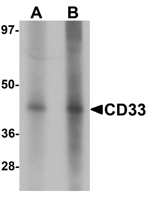
| Application Notes | CD33 antibody can be used for detection of CD33 by Western blot at 1 - 2 µg/mL. |
| Gene ID | 945 |
|---|---|
| Other Names | Myeloid cell surface antigen CD33, Sialic acid-binding Ig-like lectin 3, Siglec-3, gp67, CD33, CD33, SIGLEC3 |
| Target/Specificity | CD33; At least three isoforms of CD33 are known to exist; this antibody will detect all three isoforms. |
| Reconstitution & Storage | CD33 antibody can be stored at 4℃ for three months and -20℃, stable for up to one year. As with all antibodies care should be taken to avoid repeated freeze thaw cycles. Antibodies should not be exposed to prolonged high temperatures. |
| Precautions | CD33 Antibody is for research use only and not for use in diagnostic or therapeutic procedures. |
For Research Use Only. Not For Use In Diagnostic Procedures.
| Name | CD33 |
|---|---|
| Synonyms | SIGLEC3 |
| Function | Sialic-acid-binding immunoglobulin-like lectin (Siglec) that plays a role in mediating cell-cell interactions and in maintaining immune cells in a resting state (PubMed:10611343, PubMed:11320212, PubMed:15597323). Preferentially recognizes and binds alpha-2,3- and more avidly alpha-2,6-linked sialic acid-bearing glycans (PubMed:7718872). Upon engagement of ligands such as C1q or syalylated glycoproteins, two immunoreceptor tyrosine-based inhibitory motifs (ITIMs) located in CD33 cytoplasmic tail are phosphorylated by Src-like kinases such as LCK (PubMed:10887109, PubMed:28325905). These phosphorylations provide docking sites for the recruitment and activation of protein-tyrosine phosphatases PTPN6/SHP-1 and PTPN11/SHP- 2 (PubMed:10206955, PubMed:10556798, PubMed:10887109). In turn, these phosphatases regulate downstream pathways through dephosphorylation of signaling molecules (PubMed:10206955, PubMed:10887109). One of the repressive effect of CD33 on monocyte activation requires phosphoinositide 3-kinase/PI3K (PubMed:15597323). |
| Cellular Location | [Isoform CD33M]: Cell membrane; Single-pass type I membrane protein |
| Tissue Location | Monocytic/myeloid lineage cells. In the brain, CD33 is mainly expressed on microglial cells |
Provided below are standard protocols that you may find useful for product applications.
BACKGROUND
CD33 Antibody: CD33 is a member of the sialic acid-binding immunoglobulin-like lectin (Siglec) family that is highly expressed on myeloid progenitor cells. Assessment of CD33 expression is of great importance in the immunodiagnosis of acute leukemia, allowing distinction between myeloid and lymphoid origin, as CD33 is generally restricted to the myelomonocytic lineage. CD33 can associate with the protein-tyrosine phosphatases SHP-1 and SHP-2 and thus could modulate downstream signaling events associated with cell activation. Common variants of CD33 have been found to be associated with late-onset Alzheimer's disease.
REFERENCES
Freeman SD, Kelm S, Barber EK, et al. Characterization of CD33 as a new member of the sialoadhesion family of cellular interaction molecules. Blood 1995; 85:2005-12.
Crocker PR and Varki A. Siglecs, sialic acids, and innate immunity. Trends Immunol. 2001; 22:337-42.
Taylor VC, Buckley CD, Douglass M, et al. The myeloid-specific sialic acid-binding receptor, CD33, associates with the protein-tyrosine phosphatases, SHP-1 and SHP-2. J. Biol. Chem. 1999; 274:11505-12
Naj AC, Jun G, Beecham GW, et al. Common variants at MS4A4/MS4A6E, CD2AP, CD33 and EPHA are associated with late-onset Alzheimer’s disease. Nat. Genet. 2011; 43:436-41

终于等到您。ABCEPTA(百远生物)抗体产品。
点击下方“我要评价 ”按钮提交您的反馈信息,您的反馈和评价是我们最宝贵的财富之一,
我们将在1-3个工作日内处理您的反馈信息。
如有疑问,联系:0512-88856768 tech-china@abcepta.com.


