


 癌症的基本特征包括细胞增殖、血管生成、迁移、凋亡逃避机制和细胞永生等。找到癌症发生过程中这些通路的关键标记物和对应的抗体用于检测至关重要。
 为您推荐一个泛素化位点预测神器——泛素化分析工具,可以为您的蛋白的泛素化位点作出预测和评分。
 细胞自噬受体图形绘图工具为你的蛋白的细胞受体结合位点作出预测和评分,识别结合到自噬通路中的蛋白是非常重要的,便于让我们理解自噬在正常生理、病理过程中的作用,如发育、细胞分化、神经退化性疾病、压力条件下、感染和癌症。
Periphilin Antibody
- 产品详情
- 实验流程
- 背景知识
Application
| WB, IF, E, IHC-P |
|---|---|
| Primary Accession | Q8NEY8 |
| Other Accession | NP_958848, 48255929 |
| Reactivity | Human, Mouse |
| Host | Rabbit |
| Clonality | Polyclonal |
| Isotype | IgG |
| Calculated MW | 52737 Da |
| Concentration (mg/ml) | 1 mg/mL |
| Conjugate | Unconjugated |
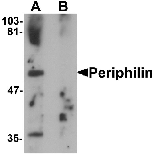
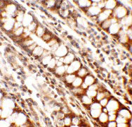
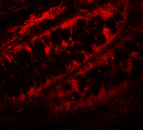
| Application Notes | Periphilin antibody can be used for detection of Periphilin by Western blot at 1 µg/mL. Antibody can also be used for immunohistochemistry starting at 2.5 µg/mL. For immunofluorescence start at 5 µg/mL. |
| Gene ID | 51535 |
|---|---|
| Other Names | Periphilin-1, Gastric cancer antigen Ga50, PPHLN1 |
| Target/Specificity | PPHLN1; |
| Reconstitution & Storage | Periphilin antibody can be stored at 4℃ for three months and -20℃, stable for up to one year. As with all antibodies care should be taken to avoid repeated freeze thaw cycles. Antibodies should not be exposed to prolonged high temperatures. |
| Precautions | Periphilin Antibody is for research use only and not for use in diagnostic or therapeutic procedures. |
| Name | PPHLN1 (HGNC:19369) |
|---|---|
| Function | Component of the HUSH complex, a multiprotein complex that mediates epigenetic repression. The HUSH complex is recruited to genomic loci rich in H3K9me3 and is probably required to maintain transcriptional silencing by promoting recruitment of SETDB1, a histone methyltransferase that mediates further deposition of H3K9me3. In the HUSH complex, contributes to the maintenance of the complex at chromatin (PubMed:26022416). Acts as a transcriptional corepressor and regulates the cell cycle, probably via the HUSH complex (PubMed:15474462, PubMed:17963697). The HUSH complex is also involved in the silencing of unintegrated retroviral DNA: some part of the retroviral DNA formed immediately after infection remains unintegrated in the host genome and is transcriptionally repressed (PubMed:30487602). May be involved in epithelial differentiation by contributing to epidermal integrity and barrier formation (PubMed:12853457). |
| Cellular Location | Nucleus. Cytoplasm. Chromosome. Note=In undifferentiated keratinocytes expressed in speckle-type nuclear granules and at the nuclear membrane, but in the differentiated keratinocytes colocalized with periplakin at the cell periphery and at cell-cell junctions (PubMed:12853457) Localizes to chromatin (PubMed:26022416). |
| Tissue Location | Ubiquitous.. |
For Research Use Only. Not For Use In Diagnostic Procedures.
Provided below are standard protocols that you may find useful for product applications.
BACKGROUND
Periphilin Antibody: Periphilin, known as PPHLN1 or gastric cancer antigen Ga50, plays a role in epithelial differentiation and contributes to epidermal integrity and barrier formation. It interacts with periplakin, a known precursor of the cornified cell envelope. Periphilin 1 is ubiquitously expressed and localizes to nucleus and cytoplasm. Existing as eight alternatively spliced isoforms, Periphilin is highly insoluble and contains a putative nuclear localization signal (NLS) within its N-terminal half, a prerequisite for the formation of insoluble complexes and a possible caspase recognition sequence and a potential nuclear export signal. Periphilin may play an important role in the nervous system.
REFERENCES
Kazerounian S and Aho S. Characterization of periphilin, a widespread, highly insoluble nuclear protein and potential constituent of the keratinocyte cornified envelope. J. Biol. Chem. 2003; 278:36707-17.
Kurita M, Suzuki H, Masai H, et al. Overexpression of CR/periphilin downregulates Cdc7 expression and induces S-phase arrest. Biochem. Biophys. Res. Commun. 2004; 324:554-61
Soehn AS, Pham TT, Schaeferhoff K, et al. Periphilin is strongly expressed in the murine nervous system and is indispensable for murine development. Genesis 2009; 47:697-707
Soehn AS, Franck T, Biskup S, et al. Periphilin is a novel interactor of synphilin-1, a protein implicated in Parkinson's disease. Neurogenetics 2010; 11:203-15
终于等到您。ABCEPTA(百远生物)抗体产品。
点击下方“我要评价 ”按钮提交您的反馈信息,您的反馈和评价是我们最宝贵的财富之一,
我们将在1-3个工作日内处理您的反馈信息。
如有疑问,联系:0512-88856768 tech-china@abcepta.com.


