


 癌症的基本特征包括细胞增殖、血管生成、迁移、凋亡逃避机制和细胞永生等。找到癌症发生过程中这些通路的关键标记物和对应的抗体用于检测至关重要。
 为您推荐一个泛素化位点预测神器——泛素化分析工具,可以为您的蛋白的泛素化位点作出预测和评分。
 细胞自噬受体图形绘图工具为你的蛋白的细胞受体结合位点作出预测和评分,识别结合到自噬通路中的蛋白是非常重要的,便于让我们理解自噬在正常生理、病理过程中的作用,如发育、细胞分化、神经退化性疾病、压力条件下、感染和癌症。
CASK Antibody

- 产品详情
- 实验流程
- 背景知识
Application
| WB, IF, E |
|---|---|
| Primary Accession | O14936 |
| Other Accession | NP_003679, 186972120 |
| Reactivity | Human, Mouse, Rat |
| Host | Rabbit |
| Clonality | Polyclonal |
| Isotype | IgG |
| Calculated MW | 105123 Da |
| Concentration (mg/ml) | 1 mg/mL |
| Conjugate | Unconjugated |
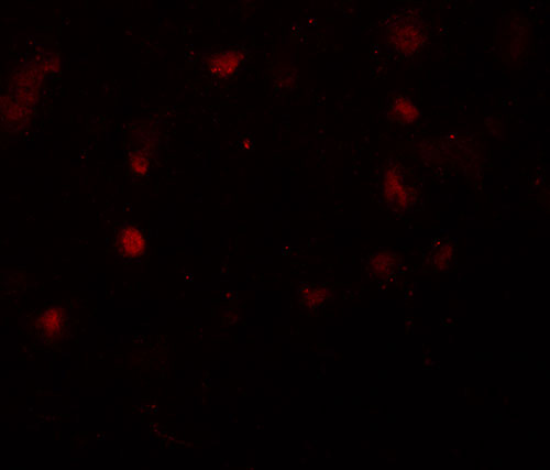
| Application Notes | CASK antibody can be used for detection of CASK by Western blot at 1 µg/mL. For immunofluorescence start at 20 µg/mL. |
| Gene ID | 8573 |
|---|---|
| Other Names | Peripheral plasma membrane protein CASK, hCASK, 2.7.11.1, Calcium/calmodulin-dependent serine protein kinase, Protein lin-2 homolog, CASK, LIN2 |
| Target/Specificity | CASK; At least six alternatively spliced transcript variants have been observed. |
| Reconstitution & Storage | CASK antibody can be stored at 4℃ for three months and -20℃, stable for up to one year. As with all antibodies care should be taken to avoid repeated freeze thaw cycles. Antibodies should not be exposed to prolonged high temperatures. |
| Precautions | CASK Antibody is for research use only and not for use in diagnostic or therapeutic procedures. |
| Name | CASK (HGNC:1497) |
|---|---|
| Synonyms | LIN2 |
| Function | Multidomain scaffolding Mg(2+)-independent protein kinase that catalyzes the phosphotransfer from ATP to proteins such as NRXN1, and plays a role in synaptic transmembrane protein anchoring and ion channel trafficking (PubMed:18423203). Contributes to neural development and regulation of gene expression via interaction with the transcription factor TBR1. Binds to cell-surface proteins, including amyloid precursor protein, neurexins and syndecans. May mediate a link between the extracellular matrix and the actin cytoskeleton via its interaction with syndecan and with the actin/spectrin-binding protein 4.1. Component of the LIN-10-LIN-2-LIN-7 complex, which associates with the motor protein KIF17 to transport vesicles containing N-methyl-D- aspartate (NMDA) receptor subunit NR2B along microtubules (By similarity). |
| Cellular Location | Nucleus {ECO:0000250|UniProtKB:Q62915}. Cytoplasm {ECO:0000250|UniProtKB:Q62915}. Cell membrane {ECO:0000250|UniProtKB:Q62915}; Peripheral membrane protein {ECO:0000250|UniProtKB:Q62915} |
| Tissue Location | Ubiquitous. Expression is significantly greater in brain relative to kidney, lung, and liver and in fetal brain and kidney relative to lung and liver. |
For Research Use Only. Not For Use In Diagnostic Procedures.
Provided below are standard protocols that you may find useful for product applications.
BACKGROUND
CASK Antibody: CASK (Calcium/calmodulin-dependent serine protein kinase), a conserved multi-domain scaffolding protein, belongs to a MAGUK (membrane-associated guanylate kinase homologs) subfamily and is involved in cell junction organization, tumor suppression and signaling. It is characterized by a novel domain structure that consists of a calcium/calmodulin-dependent protein kinase domain followed by PDZ, SH3 and guanylate kinase-like (GUK) domains. CASK is ubiquitously expressed and significantly greater in brain where it is thought to be involved in signaling at neuronal synapses. CASK interacts with CASKINs and defects in CASK are the cause of mental retardation X-linked CASK-related.
REFERENCES
Dimitratos SD, Woods DF, and Bryant PJ. Camguk, Lin-2, and CASK: novel membrane associated guanylate kinase homologs that also contain CaM kinase domains. Mech. Dev. 1997; 63:127-30.
Hsueh YP and Sheng M. Regulated expression and subcellular localization of syndecan heparan sulfate proteoglycans and the syndecan-binding protein CASK/LIN-2 during rat brain development. J. Neurosci. 1999;19:7415-25.
Tabuchi K, Biederer T, Butz S, et al. CASK participates in alternative tripartite complexes in which Mint 1 competes for binding with CASKIN2, a novel CASK-binding protein. J. Neurosci. 2002; 22:4264-73.
Kuo TY, Hong CJ, Chien HL, et al. X-linked mental retardation gene CASK interacts with Bcl11A/CTIP1 and regulates axon branching and outgrowth. J. Neurosci. Res. 2010; 88: 2364-73.
终于等到您。ABCEPTA(百远生物)抗体产品。
点击下方“我要评价 ”按钮提交您的反馈信息,您的反馈和评价是我们最宝贵的财富之一,
我们将在1-3个工作日内处理您的反馈信息。
如有疑问,联系:0512-88856768 tech-china@abcepta.com.


