


 癌症的基本特征包括细胞增殖、血管生成、迁移、凋亡逃避机制和细胞永生等。找到癌症发生过程中这些通路的关键标记物和对应的抗体用于检测至关重要。
 为您推荐一个泛素化位点预测神器——泛素化分析工具,可以为您的蛋白的泛素化位点作出预测和评分。
 细胞自噬受体图形绘图工具为你的蛋白的细胞受体结合位点作出预测和评分,识别结合到自噬通路中的蛋白是非常重要的,便于让我们理解自噬在正常生理、病理过程中的作用,如发育、细胞分化、神经退化性疾病、压力条件下、感染和癌症。
WIPI2 Antibody
- 产品详情
- 实验流程
- 背景知识
Application
| WB, E |
|---|---|
| Primary Accession | Q9Y4P8 |
| Other Accession | NP_056425, 7661580 |
| Reactivity | Human, Rat |
| Host | Rabbit |
| Clonality | Polyclonal |
| Isotype | IgG |
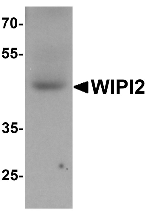
| Calculated MW | 49408 Da |
| Concentration (mg/ml) | 1 mg/mL |
| Conjugate | Unconjugated |
| Application Notes | WIPI2 antibody can be used for detection of WIPI2 by Western blot at 1 - 2 µg/mL. |
| Gene ID | 26100 |
|---|---|
| Other Names | WD repeat domain phosphoinositide-interacting protein 2, WIPI-2, WIPI49-like protein 2, WIPI2 |
| Target/Specificity | WIPI2; WIPI2 antibody is human and rat reactive. Multiple isoforms of WIPI2 are known to exist. WIPI2 antibody is predicted to not cross-react with WIPI1. |
| Reconstitution & Storage | WIPI2 antibody can be stored at 4℃ for three months and -20℃, stable for up to one year. As with all antibodies care should be taken to avoid repeated freeze thaw cycles. Antibodies should not be exposed to prolonged high temperatures. |
| Precautions | WIPI2 Antibody is for research use only and not for use in diagnostic or therapeutic procedures. |
| Name | WIPI2 (HGNC:32225) |
|---|---|
| Function | Component of the autophagy machinery that controls the major intracellular degradation process by which cytoplasmic materials are packaged into autophagosomes and delivered to lysosomes for degradation (PubMed:20505359, PubMed:28561066). Involved in an early step of the formation of preautophagosomal structures (PubMed:20505359, PubMed:28561066). Binds and is activated by phosphatidylinositol 3- phosphate (PtdIns3P) forming on membranes of the endoplasmic reticulum upon activation of the upstream ULK1 and PI3 kinases (PubMed:28561066). Mediates ER-isolation membranes contacts by interacting with the ULK1:RB1CC1 complex and PtdIns3P (PubMed:28890335). Once activated, WIPI2 recruits at phagophore assembly sites the ATG12-ATG5-ATG16L1 complex that directly controls the elongation of the nascent autophagosomal membrane (PubMed:20505359, PubMed:28561066). |
| Cellular Location | Preautophagosomal structure membrane; Peripheral membrane protein; Cytoplasmic side. Note=Localizes to omegasomes membranes which are endoplasmic reticulum connected structures at the origin of preautophagosomal structures. Enriched at preautophagosomal structure membranes in response to PtdIns3P. |
| Tissue Location | Ubiquitously expressed (at protein level). Highly expressed in heart, skeletal muscle and pancreas. Expression is down- regulated in pancreatic and in kidney tumors |
For Research Use Only. Not For Use In Diagnostic Procedures.
Provided below are standard protocols that you may find useful for product applications.
BACKGROUND
WIPI2 Antibody: WD repeat proteins play a role in many essential biologic functions, regulating the assembly of multiprotein complexes by presenting a beta-propeller platform for simultaneous and reversible protein-protein interactions. WIPI2, also known as ATG18B or ATG21, is a human homolog to yeast ATG18 and contains three WD repeats and has a 7-bladed propeller structure with a conserved motif that facilitates its interaction with other proteins. It is recruited to early autophagosomal structures along with Atg16L and ULK1 and is required for the formation of LC3-positive autophagosomes. Along with the highly related WIPI1, WIPI2 is found at the plasma membrane in addition to autophagosomal membranes.
REFERENCES
Smith TF. Diversity of WD-repeat proteins. Subcell. Biochem. 2008; 48:20-30.
Polson HE, de Lartique J, Rigden DJ, et al. Mammalian ATG18 (WIPI2) localizes to moegasome-anchored phagophores and positively regulates LC3 lipidation. Autophagy 2010; 6:506-22.
Proikas-Cezanne T and Robenek H. Freeze-fracture replica immunolabelling reveals human WIPI-1 and WIPI-2 as membrane proteins of autophagosomes. J. Cell. Mol. Med. 2011; 15:2007-10.
终于等到您。ABCEPTA(百远生物)抗体产品。
点击下方“我要评价 ”按钮提交您的反馈信息,您的反馈和评价是我们最宝贵的财富之一,
我们将在1-3个工作日内处理您的反馈信息。
如有疑问,联系:0512-88856768 tech-china@abcepta.com.


