


 癌症的基本特征包括细胞增殖、血管生成、迁移、凋亡逃避机制和细胞永生等。找到癌症发生过程中这些通路的关键标记物和对应的抗体用于检测至关重要。
 为您推荐一个泛素化位点预测神器——泛素化分析工具,可以为您的蛋白的泛素化位点作出预测和评分。
 细胞自噬受体图形绘图工具为你的蛋白的细胞受体结合位点作出预测和评分,识别结合到自噬通路中的蛋白是非常重要的,便于让我们理解自噬在正常生理、病理过程中的作用,如发育、细胞分化、神经退化性疾病、压力条件下、感染和癌症。
HEPACAM2 Antibody
- 产品详情
- 实验流程
- 背景知识
Application
| WB, E |
|---|---|
| Primary Accession | A8MVW5 |
| Other Accession | NP_001034461, 86439957 |
| Reactivity | Human, Mouse, Rat |
| Host | Rabbit |
| Clonality | Polyclonal |
| Isotype | IgG |
| Calculated MW | 51407 Da |
| Concentration (mg/ml) | 1 mg/mL |
| Conjugate | Unconjugated |
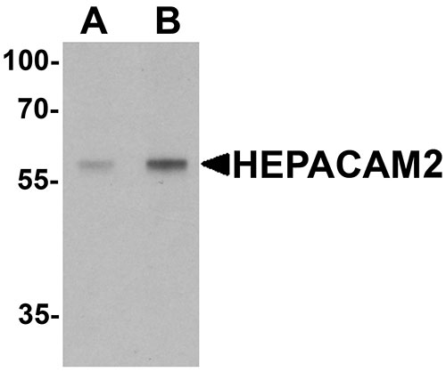
| Application Notes | HEPACAM2 antibody can be used for detection of HEPACAM2 by Western blot at 0.5 - 1 µg/mL. |
| Gene ID | 253012 |
|---|---|
| Other Names | HEPACAM family member 2, Mitotic kinetics regulator, HEPACAM2, MIKI |
| Target/Specificity | HEPACAM2; HEPACAM2 antibody is human, mouse and rat reactive. At least two isoforms of HEPACAM2 are known to exist; this antibody will detect both isoforms. HEPACAM2 antibody is predicted to not cross-react with HEPACAM. |
| Reconstitution & Storage | HEPACAM2 antibody can be stored at 4℃ for three months and -20℃, stable for up to one year. As with all antibodies care should be taken to avoid repeated freeze thaw cycles. Antibodies should not be exposed to prolonged high temperatures. |
| Precautions | HEPACAM2 Antibody is for research use only and not for use in diagnostic or therapeutic procedures. |
| Name | HEPACAM2 |
|---|---|
| Synonyms | MIKI |
| Function | Required during prometaphase for centrosome maturation. Following poly-ADP-ribosylation (PARsylation) by TNKS, translocates from the Golgi apparatus to mitotic centrosomes and plays a key role in the formation of robust microtubules for prompt movement of chromosomes: anchors AKAP9/CG-NAP, a scaffold protein of the gamma- tubulin ring complex and promotes centrosome maturation. |
| Cellular Location | Golgi apparatus membrane; Single-pass type I membrane protein. Cytoplasm, cytoskeleton, spindle. Cytoplasm, cytoskeleton, microtubule organizing center, centrosome. Midbody. Note=In interphase, localizes to the Golgi apparatus. Localizes to centrosomes and spindles during prophase, prometaphase, and metaphase of mitosis, and to midbodies at telophase Translocation to mitotic centrosomes is the result of poly-ADP- ribosylation (PARsylation). |
| Tissue Location | Widely expressed.. |
For Research Use Only. Not For Use In Diagnostic Procedures.
Provided below are standard protocols that you may find useful for product applications.
BACKGROUND
HEPACAM2 Antibody: HEPACAM2 (Hepatocyte cell adhesion molecule 2), a type I N-linked transmembrane glycoprotein, belongs to the immunoglobulin superfamily. The exact function of HEPACAM2 is currently unknown, but the related protein HEPACAM forms cishomodimers on the cell surface to regulate the cell adhesion and may inhibit cell growth through suppression of cell proliferation. HEPACAM and HEPACAM2 mRNA are differentially regulated in canine tumors, with HEPACAM2 mRNA showing increased levels in adenomas, but decreased levels in metastatic carcinomas compared to normal tissues, while HEPACAM protein levels decreased in adenomas. It is therefore likely that HEPACAM2 plays a different role than HEPACAM in the development and progression to tumors.
REFERENCES
Klopfleisch R, Klose P, da Costa A, et al. HEPACAM1 and 2 are differentially regulated in canine mammary adenomas and carcinomas and its lymph nodes metastases. BMC Veterinary Res. 2010; 6:15.
Moh MC, Tian Q, Zhang T, et al. The immunoglobulin-like cell adhesion molecule HEPACAM modulate cell adhesion and motility through direct interaction with the actin cytoskeleton. J. Cell Physiol. 2009; 219:382-91.
Lee LH, Moh MC, Zhang T, et al. The immunoglobulin-like cell adhesion molecule HEPACAM induces differentiation of human glioblastoma U373-MG cells. J. Cell Biochem. 2009; 107:1129-38.
终于等到您。ABCEPTA(百远生物)抗体产品。
点击下方“我要评价 ”按钮提交您的反馈信息,您的反馈和评价是我们最宝贵的财富之一,
我们将在1-3个工作日内处理您的反馈信息。
如有疑问,联系:0512-88856768 tech-china@abcepta.com.


