


 癌症的基本特征包括细胞增殖、血管生成、迁移、凋亡逃避机制和细胞永生等。找到癌症发生过程中这些通路的关键标记物和对应的抗体用于检测至关重要。
 为您推荐一个泛素化位点预测神器——泛素化分析工具,可以为您的蛋白的泛素化位点作出预测和评分。
 细胞自噬受体图形绘图工具为你的蛋白的细胞受体结合位点作出预测和评分,识别结合到自噬通路中的蛋白是非常重要的,便于让我们理解自噬在正常生理、病理过程中的作用,如发育、细胞分化、神经退化性疾病、压力条件下、感染和癌症。
KANK1 Antibody
- 产品详情
- 实验流程
- 背景知识
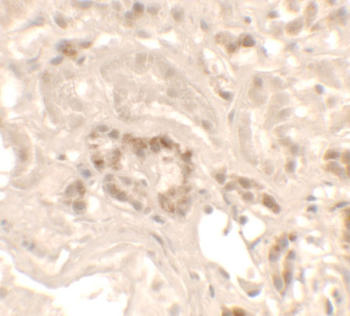
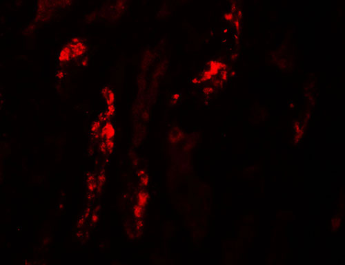
Application
| WB, IF, E, IHC-P |
|---|---|
| Primary Accession | Q14678 |
| Other Accession | NP_055973, 64464726 |
| Reactivity | Human, Mouse |
| Host | Rabbit |
| Clonality | Polyclonal |
| Isotype | IgG |
| Calculated MW | 147289 Da |
| Concentration (mg/ml) | 1 mg/mL |
| Conjugate | Unconjugated |
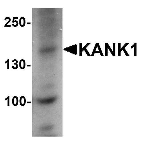
| Application Notes | KANK1 Antibody can be used for detection of KANK1 by Western blot at 1 µg/mL. |
| Gene ID | 23189 |
|---|---|
| Other Names | KN motif and ankyrin repeat domain-containing protein 1, Ankyrin repeat domain-containing protein 15, Kidney ankyrin repeat-containing protein, KANK1, ANKRD15, KANK, KIAA0172 |
| Target/Specificity | KANK1; Two alternatively spliced transcript variants encoding different isoforms have been identified. The lower molecular weight band seen in the immunoblot is thought to be non-specific. |
| Reconstitution & Storage | KANK1 antibody can be stored at 4℃ for three months and -20℃, stable for up to one year. |
| Precautions | KANK1 Antibody is for research use only and not for use in diagnostic or therapeutic procedures. |
| Name | KANK1 (HGNC:19309) |
|---|---|
| Function | Adapter protein that links structural and signaling protein complexes positioned to guide microtubule and actin cytoskeleton dynamics during cell morphogenesis (PubMed:22084092, PubMed:24120883). At focal adhesions (FAs) rims, organizes cortical microtubule stabilizing complexes (CMSCs) and directly interacts with major FA component TLN1, forming macromolecular assemblies positioned to control microtubule-actin crosstalk at the cell edge (PubMed:24120883, PubMed:27410476). Recruits KIF21A in CMSCs at axonal growth cones and regulates axon guidance by suppressing microtubule growth without inducing microtubule disassembly once it reaches the cell cortex (PubMed:24120883). Interacts with ARFGEF1 and participates in establishing microtubule-organizing center (MTOC) orientation and directed cell movement in wound healing (PubMed:22084092). Regulates actin stress fiber formation and cell migration by inhibiting RHOA activation in response to growth factors; this function involves phosphorylation through PI3K/Akt signaling and may depend on the competitive interaction with 14-3-3 adapter proteins to sequester them from active complexes (PubMed:18458160, PubMed:25961457). Inhibits the formation of lamellipodia but not of filopodia; this function may depend on the competitive interaction with BAIAP2 to block its association with activated RAC1. Inhibits fibronectin-mediated cell spreading; this function is partially mediated by BAIAP2 (PubMed:19171758). In the nucleus, is involved in beta-catenin- dependent activation of transcription (PubMed:16968744). During cell division, may regulate DAAM1-dependent RHOA activation that signals centrosome maturation and chromosomal segregation. May also be involved in contractile ring formation during cytokinesis (By similarity). Potential tumor suppressor for renal cell carcinoma (Probable). |
| Cellular Location | Cytoplasm, cell cortex. Cell projection, ruffle membrane; Peripheral membrane protein. Cytoplasm. Nucleus. Note=Shuttles between the cytoplasm and nucleus (PubMed:16968744). Colocalizes with CMSC components at focal adhesion rims. Colocalizes with KIF21A in membrane ruffles (PubMed:19559006, PubMed:27410476). Colocalizes with RHOA at the contractile ring. Colocalizes with RHOA and DAAM1 around centrosomes {ECO:0000250|UniProtKB:E9Q238, ECO:0000269|PubMed:16968744, ECO:0000269|PubMed:19559006, ECO:0000269|PubMed:27410476} [Isoform 2]: Cytoplasm. Nucleus Note=Shuttles between the cytoplasm and nucleus |
| Tissue Location | Widely expressed. Isoform 1 is predominantly expressed in heart and kidney. Isoform 2 probably is widely expressed at basic levels. |
For Research Use Only. Not For Use In Diagnostic Procedures.
Provided below are standard protocols that you may find useful for product applications.
BACKGROUND
KANK1 Antibody: Ankyrins are membrane adaptor molecules that play important roles in the control of cytoskeleton formation by regulating actin polymerization. KANK1 (KN motif and ankyrin repeat domain-containing protein 1), also known as ANKRD15, is a 1,352 amino acid protein that contains at least 12 exons and 5 ANK repeats. It binds to beta-catenin and regulates its subcellular distribution. KANK1 is ubiquitously expressed and localizes to cytoplasm. It may function as a tumor suppressor for renal cell carcinoma. Mutations in this gene cause cerebral palsy spastic quadriplegic type 2, a central nervous system development disorder.
REFERENCES
Zhu Y, Kakinuma N, Wang Y, et al. Kank proteins: a new family of ankyrin-repeat domain containing proteins. Biochim. Biophys. Acta 2008; 1780:128-33.
Roy BC, Kakinuma N, Kiyama R. Kank attenuates actin remodeling by preventing interaction between IRSp53 and Rac1. J. Cell Biol. 2009; 184:253-67.
Sarkar S, Roy BC, Hatano N, et al. A novel ankyrin repeat-containing gene (Kank) located at 9p24 is a growth suppressor of renal cell carcinoma. J. Biol. Chem. 2002; 277:36585-91.
Lerer I, Sagi M, Meiner V, et al. Deletion of the ANKRD15 gene at 9p24.3 causes parent-of-origin-dependent inheritance of familial cerebral palsy. Hum. Mol. Genet. 2005; 14: 3911-20
终于等到您。ABCEPTA(百远生物)抗体产品。
点击下方“我要评价 ”按钮提交您的反馈信息,您的反馈和评价是我们最宝贵的财富之一,
我们将在1-3个工作日内处理您的反馈信息。
如有疑问,联系:0512-88856768 tech-china@abcepta.com.


