


 癌症的基本特征包括细胞增殖、血管生成、迁移、凋亡逃避机制和细胞永生等。找到癌症发生过程中这些通路的关键标记物和对应的抗体用于检测至关重要。
 为您推荐一个泛素化位点预测神器——泛素化分析工具,可以为您的蛋白的泛素化位点作出预测和评分。
 细胞自噬受体图形绘图工具为你的蛋白的细胞受体结合位点作出预测和评分,识别结合到自噬通路中的蛋白是非常重要的,便于让我们理解自噬在正常生理、病理过程中的作用,如发育、细胞分化、神经退化性疾病、压力条件下、感染和癌症。
AKT1S1 Antibody
- 产品详情
- 实验流程
- 背景知识
Application
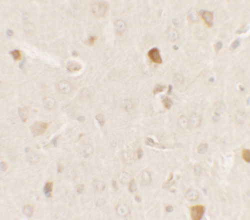
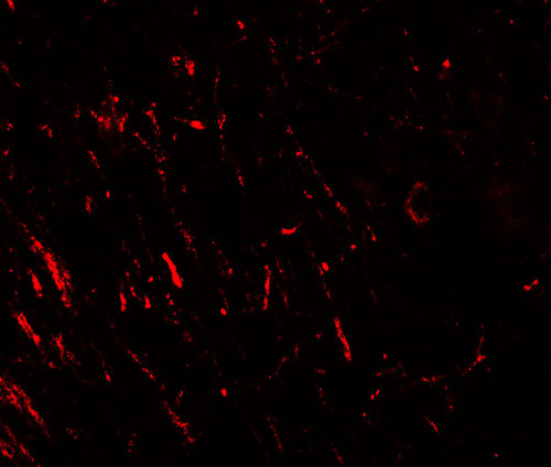
| WB, IF, E, IHC-P |
|---|---|
| Primary Accession | Q96B36 |
| Other Accession | NP_115751, 400153980 |
| Reactivity | Human, Mouse, Rat |
| Host | Rabbit |
| Clonality | Polyclonal |
| Isotype | IgG |
| Calculated MW | 27383 Da |
| Concentration (mg/ml) | 1 mg/mL |
| Conjugate | Unconjugated |
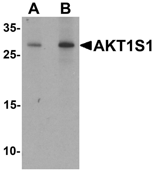
| Application Notes | AKT1S1 antibody can be used for detection of AKT1S1 by Western blot at 1 - 2 µg/mL. |
| Gene ID | 84335 |
|---|---|
| Other Names | Proline-rich AKT1 substrate 1, 40 kDa proline-rich AKT substrate, AKT1S1 {ECO:0000312|EMBL:AAH16043.1} |
| Target/Specificity | AKT1S1; AKT1S1 antibody is human, mouse and rat reactive. At least three isoforms of AKT1S1 are known to exist. |
| Reconstitution & Storage | AKT1S1 antibody can be stored at 4℃ for three months and -20℃, stable for up to one year. |
| Precautions | AKT1S1 Antibody is for research use only and not for use in diagnostic or therapeutic procedures. |
| Name | AKT1S1 {ECO:0000312|EMBL:AAH16043.1} |
|---|---|
| Function | Negative regulator of the mechanistic target of rapamycin complex 1 (mTORC1), an evolutionarily conserved central nutrient sensor that stimulates anabolic reactions and macromolecule biosynthesis to promote cellular biomass generation and growth (PubMed:17277771, PubMed:17386266, PubMed:17510057, PubMed:29236692). In absence of insulin and nutrients, AKT1S1 associates with the mTORC1 complex and directly inhibits mTORC1 activity by blocking the MTOR substrate- recruitment site (PubMed:29236692). In response to insulin and nutrients, AKT1S1 dissociates from mTORC1 (PubMed:17386266, PubMed:18372248). Its activity is dependent on its phosphorylation state and binding to 14-3-3 (PubMed:16174443, PubMed:18372248). May also play a role in nerve growth factor-mediated neuroprotection (By similarity). |
| Cellular Location | Cytoplasm, cytosol {ECO:0000250|UniProtKB:Q9D1F4}. Note=Found in the cytosolic fraction of the brain. {ECO:0000250|UniProtKB:Q9D1F4} |
| Tissue Location | Widely expressed with highest levels of expression in liver and heart. Expressed at higher levels in cancer cell lines (e.g. A-549 and HeLa) than in normal cell lines (e.g. HEK293) |
For Research Use Only. Not For Use In Diagnostic Procedures.
Provided below are standard protocols that you may find useful for product applications.
BACKGROUND
AKT1S1 Antibody: The Akt signaling pathway contributes to the regulation of apoptosis after a variety of cell death signals. AKT1S1, also known as PRAS40, is a proline-rich substrate of the kinase AKT1 and is thought to play a role in neuroprotection mediated by nerve growth factor (NGF) after transient focal cerebral ischemia (1). AKT1S1 is also a substrate and potential regulator of mammalian target of rapamycin (mTOR), a serine/threonine kinase that regulates cell growth and cell cycle, and a negative regulator of autophagy (2). Treatment with the insulin-like growth factor-1 (IGF1) can indusce the phosphorylation of AKT1S1 via the PI3K/AKT signaling pathway in PC12 cells (3).
REFERENCES
Saito A, Narasimhan P, Hayashi T, et al. Neuroprotective role of a proline-rich Akt substrate in apoptotic neuronal cell death after stroke: relationships with nerve growth factor. J. Neurosci. 2004; 24:1584-93.
Wiza C, Nascimento EB, and Ouwens DM. Role of PRAS40 in Akt and mTOR signaling in health and disease. Am. J. Physiol. Endocrinol. Metab. 2012; 302:E1453-60.
Wang H, Zhang Q, Zhang L, et al. Insulin-like growth factor-1 induces the phosphrylation of PRAS40 via the PI3K/Akt signaling pathway in PC12 cells. Neurosci. Lett. 516:105-9.
终于等到您。ABCEPTA(百远生物)抗体产品。
点击下方“我要评价 ”按钮提交您的反馈信息,您的反馈和评价是我们最宝贵的财富之一,
我们将在1-3个工作日内处理您的反馈信息。
如有疑问,联系:0512-88856768 tech-china@abcepta.com.


