


 癌症的基本特征包括细胞增殖、血管生成、迁移、凋亡逃避机制和细胞永生等。找到癌症发生过程中这些通路的关键标记物和对应的抗体用于检测至关重要。
 为您推荐一个泛素化位点预测神器——泛素化分析工具,可以为您的蛋白的泛素化位点作出预测和评分。
 细胞自噬受体图形绘图工具为你的蛋白的细胞受体结合位点作出预测和评分,识别结合到自噬通路中的蛋白是非常重要的,便于让我们理解自噬在正常生理、病理过程中的作用,如发育、细胞分化、神经退化性疾病、压力条件下、感染和癌症。
IL-36A Antibody
- 产品详情
- 实验流程
- 背景知识
Application
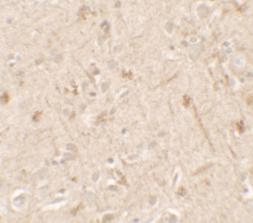
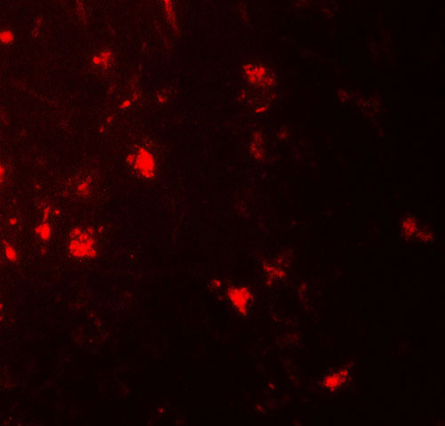
| WB, IF, E, IHC-P |
|---|---|
| Primary Accession | Q9UHA7 |
| Other Accession | NP_055255, 7657092 |
| Reactivity | Human |
| Host | Rabbit |
| Clonality | Polyclonal |
| Isotype | IgG |
| Calculated MW | 17684 Da |
| Concentration (mg/ml) | 1 mg/mL |
| Conjugate | Unconjugated |
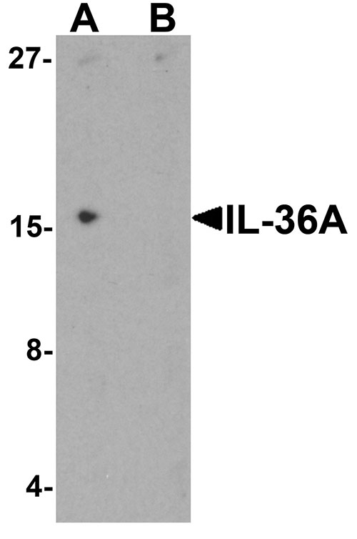
| Application Notes | IL-36A antibody can be used for detection of IL-36A by Western blot at 1 - 2 µg/ml. |
| Gene ID | 27179 |
|---|---|
| Other Names | Interleukin-36 alpha, FIL1 epsilon, Interleukin-1 epsilon, IL-1 epsilon, Interleukin-1 family member 6, IL-1F6, IL36A, FIL1E, IL1E, IL1F6 |
| Target/Specificity | IL36A; IL-36A antibody is human specific. IL-36A antibody will not cross-react with IL-36B or IL-36G. |
| Reconstitution & Storage | IL-36A antibody can be stored at 4℃ for three months and -20℃, stable for up to one year. |
| Precautions | IL-36A Antibody is for research use only and not for use in diagnostic or therapeutic procedures. |
| Name | IL36A (HGNC:15562) |
|---|---|
| Synonyms | FIL1E, IL1E, IL1F6 |
| Function | Cytokine that binds to and signals through the IL1RL2/IL-36R receptor which in turn activates NF-kappa-B and MAPK signaling pathways in target cells linked to a pro-inflammatory response. Part of the IL- 36 signaling system that is thought to be present in epithelial barriers and to take part in local inflammatory response; similar to the IL-1 system with which it shares the coreceptor IL1RAP. Seems to be involved in skin inflammatory response by acting on keratinocytes, dendritic cells and indirectly on T-cells to drive tissue infiltration, cell maturation and cell proliferation. In cultured keratinocytes induces the expression of macrophage, T-cell, and neutrophil chemokines, such as CCL3, CCL4, CCL5, CCL2, CCL17, CCL22, CL20, CCL5, CCL2, CCL17, CCL22, CXCL8, CCL20 and CXCL1, and the production of pro- inflammatory cytokines such as TNF-alpha, IL-8 and IL-6. In cultured monocytes up-regulates expression of IL-1A, IL-1B and IL-6. In myeloid dendritic cells involved in cell maturation by up-regulating surface expression of CD83, CD86 and HLA-DR. In monocyte-derived dendritic cells facilitates dendritic cell maturation and drives T-cell proliferation. May play a role in pro-inflammatory effects in the lung. |
| Cellular Location | Cytoplasm. Secreted. Note=The secretion is dependent on protein unfolding and facilitated by the cargo receptor TMED10; it results in protein translocation from the cytoplasm into the ERGIC (endoplasmic reticulum-Golgi intermediate compartment) followed by vesicle entry and secretion. |
| Tissue Location | Expressed in immune system and fetal brain, but not in other tissues tested or in multiple hematopoietic cell lines Predominantly expressed in skin keratinocytes but not in fibroblasts, endothelial cells or melanocytes. Increased in lesional psoriasis skin |
For Research Use Only. Not For Use In Diagnostic Procedures.
Provided below are standard protocols that you may find useful for product applications.
BACKGROUND
IL-36A is is a member of the interleukin 1 cytokine family whose gene and eight other interleukin 1 family genes form a cytokine gene cluster on chromosome 2 (1). IL-36A is thought to activate the NF-kappaB pathway through IL-1 receptor family members IL-1RL2 and IL-1RAcP (2). Like the related proteins IL-36B and IL-36G, IL-36A requires post-translational processing for full agonist activity, but the cleavage mechanism is currently unknown (3). The IL-36 cytokines have been suggested to amplify Th1 responses by enhancing proliferation and Th1 polarization of naive CD4+ T cells (4).
REFERENCES
Smith DE, Renshaw BR, Ketchem RR, et al. Four new members expand the interleukin-1 superfamily. J. Biol. Chem. 2000; 275:1169-75.
Towne JE, Garka KE, Renshaw BR, et al. Interleukin (IL)-1F6, IL-1F8, and IL-1F9 signal through IL-1Rrp2 and IL-1RAcP to activate the pathway leading to NF-kappaB and MAPKs. J. Biol. Chem. 2004; 279:13677-88.
Towne JE, Renshaw BR, Douangpanya J, et al. Interleukin-36 (IL-36) ligands require processing for full agonist agonist (IL-36a, IL-36b, and IL-36g) or antagonist (IL-36Ra) activity. J. Biol. Chem. 2011; 286:42594-602.
Vigne S, Palmer G, Martin P, et al. IL-36 signaling amplifies Th1 responses by enhancing proliferation and Th1 polarization of naive CD4+ T cells. Blood 2012; 120:3478-87.
终于等到您。ABCEPTA(百远生物)抗体产品。
点击下方“我要评价 ”按钮提交您的反馈信息,您的反馈和评价是我们最宝贵的财富之一,
我们将在1-3个工作日内处理您的反馈信息。
如有疑问,联系:0512-88856768 tech-china@abcepta.com.


