


 癌症的基本特征包括细胞增殖、血管生成、迁移、凋亡逃避机制和细胞永生等。找到癌症发生过程中这些通路的关键标记物和对应的抗体用于检测至关重要。
 为您推荐一个泛素化位点预测神器——泛素化分析工具,可以为您的蛋白的泛素化位点作出预测和评分。
 细胞自噬受体图形绘图工具为你的蛋白的细胞受体结合位点作出预测和评分,识别结合到自噬通路中的蛋白是非常重要的,便于让我们理解自噬在正常生理、病理过程中的作用,如发育、细胞分化、神经退化性疾病、压力条件下、感染和癌症。
TMIGD2 Antibody
- 产品详情
- 实验流程
- 背景知识
Application
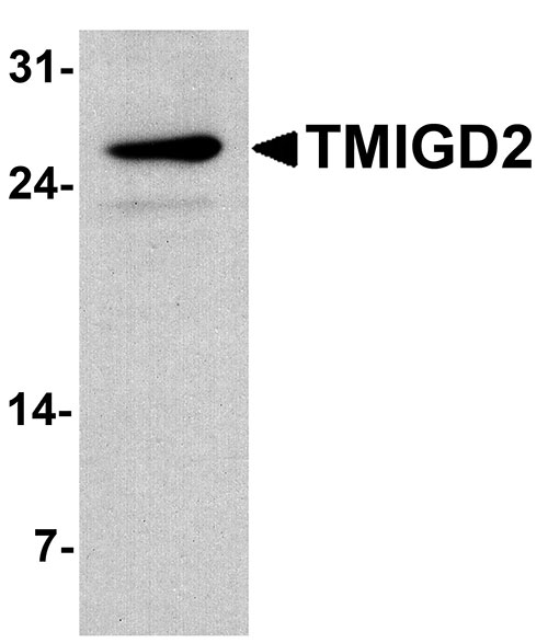
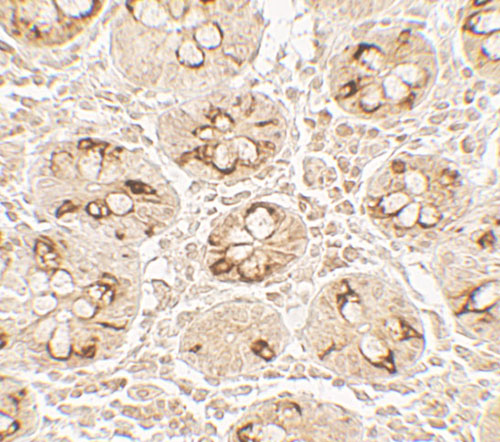
| WB, E, IHC-P |
|---|---|
| Primary Accession | Q96BF3 |
| Other Accession | NP_653216, 281306838 |
| Reactivity | Human, Mouse |
| Host | Rabbit |
| Clonality | Polyclonal |
| Isotype | IgG |
| Calculated MW | 30675 Da |
| Concentration (mg/ml) | 1 mg/mL |
| Conjugate | Unconjugated |
| Application Notes | TMIGD2 antibody can be used for detection of TMIGD2 by Western blot at 1 - 2 µg/ml. Antibody can also be used for Immunohistochemistry starting at 5 µg/mL. |
| Gene ID | 126259 |
|---|---|
| Other Names | Transmembrane and immunoglobulin domain-containing protein 2, CD28 homolog, Immunoglobulin and proline-rich receptor 1, IGPR-1, TMIGD2, CD28H, IGPR1 |
| Target/Specificity | TMIGD2; TMIGD2 antibody is human and mouse reactive. At least three isoforms of TMIGD2 are known to exist; this antibody will detect all three. TMIGD2 antibody is predicted to not cross-react with TMIGD1. |
| Reconstitution & Storage | TMIGD2 antibody can be stored at 4℃ for three months and -20℃, stable for up to one year. |
| Precautions | TMIGD2 Antibody is for research use only and not for use in diagnostic or therapeutic procedures. |
| Name | TMIGD2 |
|---|---|
| Synonyms | CD28H, IGPR1 |
| Function | Plays a role in cell-cell interaction, cell migration, and angiogenesis. Through interaction with HHLA2, costimulates T-cells in the context of TCR-mediated activation. Enhances T-cell proliferation and cytokine production via an AKT-dependent signaling cascade. |
| Cellular Location | Cell membrane; Single-pass type I membrane protein |
| Tissue Location | Widely expressed, mainly by epithelial and endothelial cells, including bronchial epithelial cells of lung, breast glandular and lobular epithelia cells, urothelium of the bladder, skin epidermis, epithelium of gastrointestinal, rectum, endometrial glands of the uterus, ureter, fallopian tube epithelium, colonic epithelium, small bowl epithelium, stomach epithelium, including both chief and parietal cells, trophoblastic epithelium of placenta, and pancreatic acinar cells (at protein level). Consistently expressed in veins and arteries (at protein level). Not detected in thyroid, cerebellum, cerebral cortex and thymus (at protein level). Expressed in lymphoid organs, with highest levels in thymus, spleen, peripheral blood lymphocytes and liver. In the thymus, expressed in CD4+ and CD8+ single- and double-positive cells, but not in immature CD4- and CD8- double-negative cells (at protein level). In peripheral blood mononuclear cells, highly expressed on CD56+ or CD16+ natural killer cells and CD3+ T-cells(at protein level). Not detected on B-cells(at protein level). Expressed in tonsils (at protein level) |
For Research Use Only. Not For Use In Diagnostic Procedures.
Provided below are standard protocols that you may find useful for product applications.
BACKGROUND
TMIGD2 (transmembrane and immunoglobulin domain containing 1), also known as immunoglobulin-containing and proline-rich receptor 1 (IGPR1), is novel adhesion molecule that is expressed in multiple tissues, primarily in cells of epithelium and endothelium origins (1). TMIGD2 is thought to be involved in angiogenesis and regulates cellular morphology, homophilic cell aggregation, and cell-cell interaction. TMIGD2 activity also modulates actin stress fiber formation and focal adhesion and reduces cell migration (1).
REFERENCES
Rahimi N, Rezazadeh K, Mahoney JE, et al. Identification of IGPR-1 as a novel adhesion molecule involved in angiogenesis. Mol. Biol. Cell 2012; 23:1646-56.
终于等到您。ABCEPTA(百远生物)抗体产品。
点击下方“我要评价 ”按钮提交您的反馈信息,您的反馈和评价是我们最宝贵的财富之一,
我们将在1-3个工作日内处理您的反馈信息。
如有疑问,联系:0512-88856768 tech-china@abcepta.com.


