


 癌症的基本特征包括细胞增殖、血管生成、迁移、凋亡逃避机制和细胞永生等。找到癌症发生过程中这些通路的关键标记物和对应的抗体用于检测至关重要。
 为您推荐一个泛素化位点预测神器——泛素化分析工具,可以为您的蛋白的泛素化位点作出预测和评分。
 细胞自噬受体图形绘图工具为你的蛋白的细胞受体结合位点作出预测和评分,识别结合到自噬通路中的蛋白是非常重要的,便于让我们理解自噬在正常生理、病理过程中的作用,如发育、细胞分化、神经退化性疾病、压力条件下、感染和癌症。
AQP2 Antibody
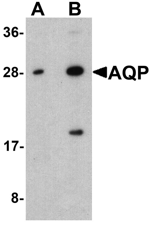
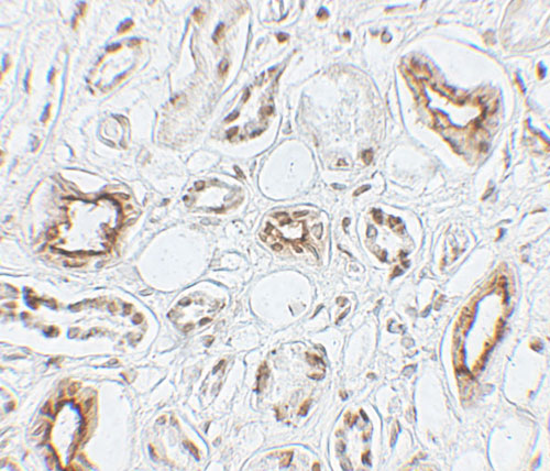
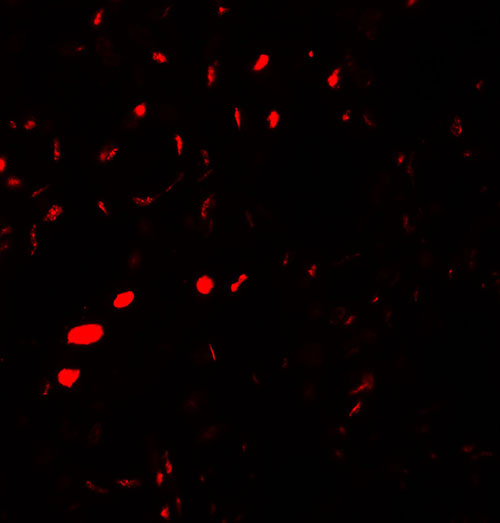
- 产品详情
- 实验流程
- 背景知识
Application 
| WB, IF, E, IHC-P |
|---|---|
| Primary Accession | P41181 |
| Other Accession | NP_000477, 4502179 |
| Reactivity | Human, Mouse, Rat |
| Host | Rabbit |
| Clonality | Polyclonal |
| Isotype | IgG |
| Calculated MW | 28837 Da |
| Concentration (mg/ml) | 1 mg/mL |
| Conjugate | Unconjugated |
| Application Notes | AQP2 antibody can be used for detection of AQP2 by Western blot at 1 - 2 µg/ml. Antibody can also be used for Immunohistochemistry starting at 5 µg/mL. For immunofluorescence start at 20 µg/mL. |
| Gene ID | 359 |
|---|---|
| Other Names | Aquaporin-2, AQP-2, ADH water channel, Aquaporin-CD, AQP-CD, Collecting duct water channel protein, WCH-CD, Water channel protein for renal collecting duct, AQP2 |
| Target/Specificity | AQP2; AQP2 antibody is human, mouse and rat reactive. This antibody is predicted to not cross-react with other members of the aquaporin protein family. |
| Reconstitution & Storage | AQP2 antibody can be stored at 4℃ for three months and -20℃, stable for up to one year. |
| Precautions | AQP2 Antibody is for research use only and not for use in diagnostic or therapeutic procedures. |
For Research Use Only. Not For Use In Diagnostic Procedures.
| Name | AQP2 (HGNC:634) |
|---|---|
| Function | Forms a water-specific channel that provides the plasma membranes of renal collecting duct with high permeability to water, thereby permitting water to move in the direction of an osmotic gradient (PubMed:15509592, PubMed:7510718, PubMed:7524315, PubMed:8140421, PubMed:8584435). Plays an essential role in renal water homeostasis (PubMed:15509592, PubMed:7524315, PubMed:8140421). Could also be permeable to glycerol (PubMed:8584435). |
| Cellular Location | Apical cell membrane; Multi-pass membrane protein. Basolateral cell membrane {ECO:0000250|UniProtKB:P34080}; Multi-pass membrane protein. Cell membrane; Multi-pass membrane protein. Cytoplasmic vesicle membrane; Multi-pass membrane protein. Golgi apparatus, trans-Golgi network membrane; Multi-pass membrane protein. Note=Shuttles from vesicles to the apical membrane (PubMed:15509592). Vasopressin-regulated phosphorylation is required for translocation to the apical cell membrane (PubMed:15509592). PLEKHA8/FAPP2 is required to transport AQP2 from the TGN to sites where AQP2 is phosphorylated (By similarity) {ECO:0000250|UniProtKB:P34080, ECO:0000269|PubMed:15509592} |
| Tissue Location | Expressed in collecting tubules in kidney medulla (at protein level) (PubMed:7510718). Detected in kidney (PubMed:7510718). |
Provided below are standard protocols that you may find useful for product applications.
BACKGROUND
Aquaporins are membrane proteins that serve in the transfer of water and small solutes across cellular membranes. One such aquaporin, aquaporin-2 (AQP2) is located in the kidney collecting tubule and plays a critical role in water reabsorbtion (1). AQP2 is mainly localized in intracellular vesicles but upon stimulation with anti-diuretic hormone (ADH), AQP2 is translocated to the apical plasma membrane by exocytic fusion of AQP2-bearing vesicles (2). Mutations in this gene have been linked to autosomal dominant and recessive forms of nephrogenic diabetes insipidus (3).
REFERENCES
Denker BM, Smith BL, Kuhada FP, et al. Identification, purification, and partial characterization of a novel Mr 28,000 integral membrane protein from erythrocytes and renal tubules. J. Biol. Chem. 1988; 263:15634-42.
Barile M, Pisitkun T, Yu MJ, et al. Large scale protein identification in intracellular aquaporin-2 vesicles from renal inner medullary collecting duct. Mol. Cell Proteomics 2005; 4:1095-106.
Bockenhauer D and Bichet DG. Inherited secondary nephrogenic diabetes insipidus: concentrating on humans. Am. J. Physiol. Renal Physiol. 2013; 304:F1037-42.

终于等到您。ABCEPTA(百远生物)抗体产品。
点击下方“我要评价 ”按钮提交您的反馈信息,您的反馈和评价是我们最宝贵的财富之一,
我们将在1-3个工作日内处理您的反馈信息。
如有疑问,联系:0512-88856768 tech-china@abcepta.com.


