


 癌症的基本特征包括细胞增殖、血管生成、迁移、凋亡逃避机制和细胞永生等。找到癌症发生过程中这些通路的关键标记物和对应的抗体用于检测至关重要。
 为您推荐一个泛素化位点预测神器——泛素化分析工具,可以为您的蛋白的泛素化位点作出预测和评分。
 细胞自噬受体图形绘图工具为你的蛋白的细胞受体结合位点作出预测和评分,识别结合到自噬通路中的蛋白是非常重要的,便于让我们理解自噬在正常生理、病理过程中的作用,如发育、细胞分化、神经退化性疾病、压力条件下、感染和癌症。
TET1 Antibody
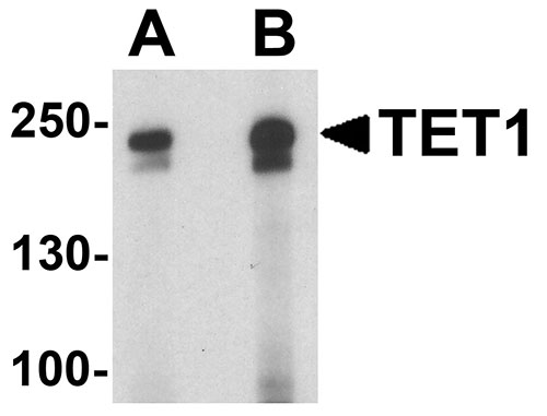
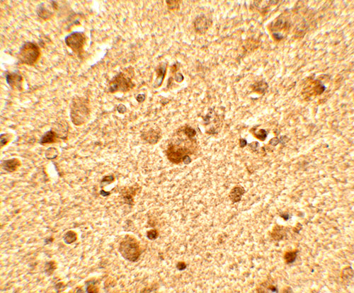
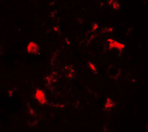
- 产品详情
- 实验流程
- 背景知识
Application 
| WB, IF, E, IHC-P |
|---|---|
| Primary Accession | Q8NFU7 |
| Other Accession | NP_085128, 156139122 |
| Reactivity | Human, Mouse, Rat |
| Host | Rabbit |
| Clonality | Polyclonal |
| Isotype | IgG |
| Calculated MW | 235309 Da |
| Concentration (mg/ml) | 1 mg/mL |
| Conjugate | Unconjugated |
| Application Notes | TET1 antibody can be used for detection of TET1 by Western blot at 1 - 2 µg/ml. Antibody can also be used for Immunohistochemistry at 5 µg/mL. For Immunoflorescence start at 20 µg/mL. |
| Gene ID | 80312 |
|---|---|
| Other Names | Methylcytosine dioxygenase TET1, 1.14.11.n2, CXXC-type zinc finger protein 6, Leukemia-associated protein with a CXXC domain, Ten-eleven translocation 1 gene protein, TET1, CXXC6, KIAA1676, LCX |
| Target/Specificity | TET1; TET1 antibody is human, mouse and rat reactive. This antibody is predicted to not cross-react with TET2 and TET3. |
| Reconstitution & Storage | TET1 antibody can be stored at 4℃ for three months and -20℃, stable for up to one year. |
| Precautions | TET1 Antibody is for research use only and not for use in diagnostic or therapeutic procedures. |
For Research Use Only. Not For Use In Diagnostic Procedures.
| Name | TET1 {ECO:0000303|PubMed:28397838, ECO:0000312|HGNC:HGNC:29484} |
|---|---|
| Function | Dioxygenase that plays a key role in active DNA demethylation, by catalyzing the sequential oxidation of the modified genomic base 5-methylcytosine (5mC) into 5-hydroxymethylcytosine (5hmC), 5-formylcytosine (5fC), and 5-carboxylcytosine (5caC) (PubMed:19372391, PubMed:21496894, PubMed:21778364, PubMed:35798741). In addition to its role in DNA demethylation, plays a more general role in chromatin regulation by recruiting histone modifying protein complexes to alter histone marks and chromatin accessibility, leading to both activation and repression of gene expression (PubMed:33833093). Plays therefore a role in many biological processes, including stem cell maintenance, T- and B-cell development, inflammation regulation, genomic imprinting, neural activity or DNA repair (PubMed:31278917). Involved in the balance between pluripotency and lineage commitment of cells and plays a role in embryonic stem cells maintenance and inner cell mass cell specification. Together with QSER1, plays an essential role in the protection and maintenance of transcriptional and developmental programs to inhibit the binding of DNMT3A/3B and therefore de novo methylation (PubMed:33833093). May play a role in pancreatic beta-cell specification during development. In this context, may function as an upstream epigenetic regulator of PAX4 presumably through direct recruitment by FOXA2 to a PAX4 enhancer to preserve its unmethylated status, thereby potentiating PAX4 expression to adopt beta-cell fate during endocrine lineage commitment (PubMed:35798741). Under DNA hypomethylation conditions, such as in female meiotic germ cells, may induce epigenetic reprogramming of pericentromeric heterochromatin (PCH), the constitutive heterochromatin of pericentromeric regions. PCH forms chromocenters in the interphase nucleus and chromocenters cluster at the prophase of meiosis. In this context, may also be essential for chromocenter clustering in a catalytic activity-independent manner, possibly through the recruitment polycomb repressive complex 1 (PRC1) to the chromocenters (By similarity). During embryonic development, may be required for normal meiotic progression in oocytes and meiotic gene activation (By similarity). Binds preferentially to DNA containing cytidine-phosphate- guanosine (CpG) dinucleotides over CpH (H=A, T, and C), hemimethylated- CpG and hemimethylated-hydroxymethyl-CpG (PubMed:29276034). |
| Cellular Location | Nucleus {ECO:0000250|UniProtKB:Q3URK3}. Chromosome. Note=Localization to chromatin is promoted by monoubiquitination on Lys-1589 [Isoform 2]: Nucleus. Chromosome {ECO:0000250|UniProtKB:Q3URK3}. Note=During DNA replication, localizes to sites of ongoing DNA replication in heterochromatin (in late S phase) in an UHRF1- and CRL4(VprBP)-dependent manner, as a consequence of ubiquitination of the conserved residue Lys-1589. Localization to heterochromatin is independent of catalytic activity |
| Tissue Location | Expressed in fetal heart, lung and brain, and in adult skeletal muscle, thymus and ovary. Not detected in adult heart, lung or brain. Up-regulated in glioblastoma cells (at protein level) (PubMed:25284789). |
Provided below are standard protocols that you may find useful for product applications.
BACKGROUND
TET1, a member of the ten-eleven-translocation (TET) family of genes, was initially discovered as a fusion partner of MLL in acute myeloid leukemias containing the t(10;11)(q22;q23) (1). It is a methylcytosine dioxygenase that catalyzes the conversion of methylcytosine to 5-hydroxymethylcytosine (2). TET1 expression is regulated during inflammation in both THP-1 and primary dendritic cells, and is a negative transcriptional regulator of IL-1beta following and inflammatory stimulus (3). Recent evidence has shown that TET1 is part of a signaling pathway that regulates breast cancer growth and metastasis (4).
REFERENCES
Lorsbach RB, Moore J, Mathew S, et al. TET1, a member of a novel protein family, is fused to MLL in acute myeloid leukemia containing the t(10;11)(q22;q23). Leukemia 2003; 17:637-41.
Tahilani M, Koh KP, Shen Y, et al. Conversion of 5-methylcytosine to 5-hydroxymethylcytosine in mammalian DNA by MLL partner TET1. Science 2009; 324:930-5.
Neves-Costa A and Moita LF. TET1 is a negative transcriptional regulator of IL-1b in the THP-1 cell line. Mol. Immunol. 2013; 54:264-70.
Sun M, Song CX, Huang H, et al. HMGA2/TET1/HOXA9 signaling pathway regulates breast cancer growth and metastasis. Proc. Natl. Acad. Sci. USA 2013; 110:9920-5.

终于等到您。ABCEPTA(百远生物)抗体产品。
点击下方“我要评价 ”按钮提交您的反馈信息,您的反馈和评价是我们最宝贵的财富之一,
我们将在1-3个工作日内处理您的反馈信息。
如有疑问,联系:0512-88856768 tech-china@abcepta.com.


