


 癌症的基本特征包括细胞增殖、血管生成、迁移、凋亡逃避机制和细胞永生等。找到癌症发生过程中这些通路的关键标记物和对应的抗体用于检测至关重要。
 为您推荐一个泛素化位点预测神器——泛素化分析工具,可以为您的蛋白的泛素化位点作出预测和评分。
 细胞自噬受体图形绘图工具为你的蛋白的细胞受体结合位点作出预测和评分,识别结合到自噬通路中的蛋白是非常重要的,便于让我们理解自噬在正常生理、病理过程中的作用,如发育、细胞分化、神经退化性疾病、压力条件下、感染和癌症。
OLIG2 Antibody

- 产品详情
- 实验流程
- 背景知识
Application
| WB, IF, E, IHC-P |
|---|---|
| Primary Accession | Q13516 |
| Other Accession | NP_005797, 17978475 |
| Reactivity | Human, Mouse, Rat |
| Host | Rabbit |
| Clonality | Polyclonal |
| Isotype | IgG |
| Calculated MW | 32385 Da |
| Concentration (mg/ml) | 1 mg/mL |
| Conjugate | Unconjugated |
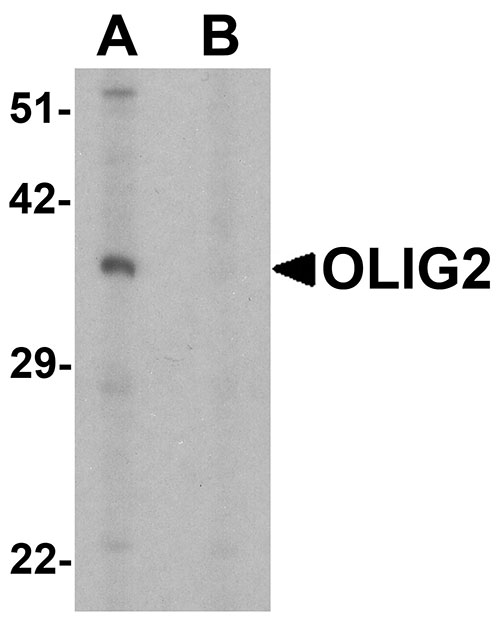
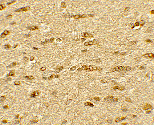
| Application Notes | OLIG2 antibody can be used for detection of OLIG2 by Western blot at 1 - 2 µg/ml. Antibody can also be used for Immunohistochemistry at 5 µg/mL. For Immunoflorescence start at 20 µg/mL. |
| Gene ID | 10215 |
|---|---|
| Other Names | Oligodendrocyte transcription factor 2, Oligo2, Class B basic helix-loop-helix protein 1, bHLHb1, Class E basic helix-loop-helix protein 19, bHLHe19, Protein kinase C-binding protein 2, Protein kinase C-binding protein RACK17, OLIG2, BHLHB1, BHLHE19, PRKCBP2, RACK17 |
| Target/Specificity | OLIG2; OLIG2 antibody is human, mouse and rat reactive. It is predicted to not cross-react with other members of the OLIG family of proteins. At least three isoforms of OLIG2 are known to exist; this antibody will detect all three isoforms. |
| Reconstitution & Storage | OLIG2 antibody can be stored at 4℃ for three months and -20℃, stable for up to one year. |
| Precautions | OLIG2 Antibody is for research use only and not for use in diagnostic or therapeutic procedures. |
| Name | OLIG2 |
|---|---|
| Synonyms | BHLHB1, BHLHE19, PRKCBP2, RACK17 |
| Function | Required for oligodendrocyte and motor neuron specification in the spinal cord, as well as for the development of somatic motor neurons in the hindbrain. Functions together with ZNF488 to promote oligodendrocyte differentiation. Cooperates with OLIG1 to establish the pMN domain of the embryonic neural tube. Antagonist of V2 interneuron and of NKX2-2-induced V3 interneuron development. |
| Cellular Location | Nucleus {ECO:0000255|PROSITE-ProRule:PRU00981}. Cytoplasm. Note=The NLS contained in the bHLH domain could be masked in the native form and translocation to the nucleus could be mediated by interaction either with class E bHLH partner protein or with NKX2-2. |
| Tissue Location | Expressed in the brain, in oligodendrocytes. Strongly expressed in oligodendrogliomas, while expression is weak to moderate in astrocytomas. Expression in glioblastomas highly variable |
For Research Use Only. Not For Use In Diagnostic Procedures.
Provided below are standard protocols that you may find useful for product applications.
BACKGROUND
The oligodendrocyte transcription factors 1 and 2 (OLIG1 and OLIG2, respectively) make up part of basic helix-loop-helix (bHLH) family of transcription factors that are specifically expressed in zones of the neuroepithelium from which oligodendrocyte precursors emerge (1). Both OLIG1 and OLIG2 genes are downstream targets of Sonic hedgehog and are expressed exclusively in the central nervous system (2). OLIG2 is first observed in the ventral most p3 progenitor domain of the ventral neural tube while OLIG1 is first expressed in the dorsal portion of the p3 domain (2). Mice overexpressing OLIG2 exhibit impaired potassium channel expression in neural progenitors and proliferation of these cells similar to that seen in Down Syndrome, suggesting that OLIG2 may play a role in this pathology (3).
REFERENCES
Zhou Q, Wang S, and Anderson DJ. Identification of a novel family of oligodendrocyte lineage-specific basic helix-loop-helix transcription factors. Neuron 2000; 25:331-43.
Lu QR, Yuk D, Alberta JA, et al. Sonic Hedgehog-regulated oligodendrocyte lineage genes encoding bHLH proteins in the mammalian central nervous system. Neuron 2000; 25:317-29.
Lu J, Lian G, Zhou H, et al. OLIG2 over-expression impairs proliferation of human Down syndrome neural progenitors. Hum. Mol. Genet. 2012; 21:2330-40.
终于等到您。ABCEPTA(百远生物)抗体产品。
点击下方“我要评价 ”按钮提交您的反馈信息,您的反馈和评价是我们最宝贵的财富之一,
我们将在1-3个工作日内处理您的反馈信息。
如有疑问,联系:0512-88856768 tech-china@abcepta.com.


