


 癌症的基本特征包括细胞增殖、血管生成、迁移、凋亡逃避机制和细胞永生等。找到癌症发生过程中这些通路的关键标记物和对应的抗体用于检测至关重要。
 为您推荐一个泛素化位点预测神器——泛素化分析工具,可以为您的蛋白的泛素化位点作出预测和评分。
 细胞自噬受体图形绘图工具为你的蛋白的细胞受体结合位点作出预测和评分,识别结合到自噬通路中的蛋白是非常重要的,便于让我们理解自噬在正常生理、病理过程中的作用,如发育、细胞分化、神经退化性疾病、压力条件下、感染和癌症。
DOCK8 Antibody
- 产品详情
- 实验流程
- 背景知识
Application
| WB, E, IHC-P |
|---|---|
| Primary Accession | Q8NF50 |
| Other Accession | NP_982272, 238231392 |
| Reactivity | Human, Mouse, Rat |
| Host | Rabbit |
| Clonality | Polyclonal |
| Isotype | IgG |
| Calculated MW | 238529 Da |
| Concentration (mg/ml) | 1 mg/mL |
| Conjugate | Unconjugated |
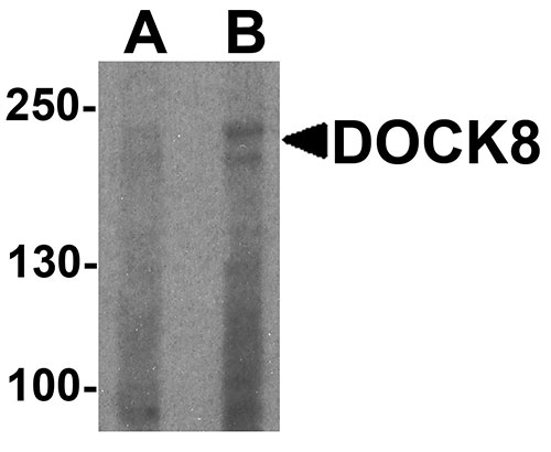
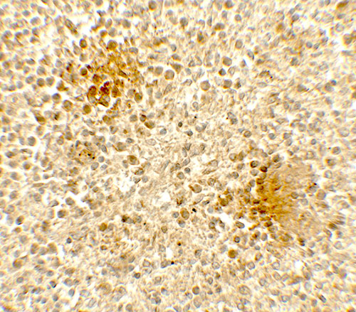
| Application Notes | DOCK8 antibody can be used for detection of DOCK8 by Western blot at 1 - 2 µg/ml. Antibody can also be used for Immunohistochemistry at 5 µg/mL. |
| Gene ID | 81704 |
|---|---|
| Other Names | Dedicator of cytokinesis protein 8, DOCK8 |
| Target/Specificity | DOCK8; DOCK8 antibody is human, mouse and rat reactive. Multiple isoforms of DOCK8 are known to exist. |
| Reconstitution & Storage | DOCK8 antibody can be stored at 4℃ for three months and -20℃, stable for up to one year. |
| Precautions | DOCK8 Antibody is for research use only and not for use in diagnostic or therapeutic procedures. |
| Name | DOCK8 |
|---|---|
| Function | Guanine nucleotide exchange factor (GEF) which specifically activates small GTPase CDC42 by exchanging bound GDP for free GTP (PubMed:22461490, PubMed:28028151). During immune responses, required for interstitial dendritic cell (DC) migration by locally activating CDC42 at the leading edge membrane of DC (By similarity). Required for CD4(+) T-cell migration in response to chemokine stimulation by promoting CDC42 activation at T cell leading edge membrane (PubMed:28028151). Is involved in NK cell cytotoxicity by controlling polarization of microtubule-organizing center (MTOC), and possibly regulating CCDC88B-mediated lytic granule transport to MTOC during cell killing (PubMed:25762780). |
| Cellular Location | Cytoplasm. Cell membrane; Peripheral membrane protein; Cytoplasmic side. Cell projection, lamellipodium membrane; Peripheral membrane protein; Cytoplasmic side. Note=Enriched and co-localizes with GTPase CDC42 at the immunological synapse formed during T cell/antigen presenting cell cognate interaction. Translocates from the cytoplasm to the plasma membrane in response to chemokine CXCL12/SDF-1-alpha stimulation |
| Tissue Location | Expressed in peripheral blood mononuclear cells (PBMCs). |
For Research Use Only. Not For Use In Diagnostic Procedures.
Provided below are standard protocols that you may find useful for product applications.
BACKGROUND
The Dedicator of cytokinesis protein 8 (DOCK8) is a member of the DOCK180 family of guanine nucleotide exchange factors (1). DOCK8 plays an essential role in humoral immune responses and is important in the proper formation of the B cell immunological synapse (reviewed in 2). Mutations in this gene result in the autosomal recessive form of the hyper-IgE syndrome (3).
REFERENCES
Ruusala A and Aspenstrom P. Isolation and characterisation of DOCK8, a member of the DOCK180-related regulators of cell morphology. FEBS Lett. 2004; 572:159-66.
Randall KL, Lambe T, Goodnow CC, et al. The essential role of DOCK8 in humoral immunity. Dis. Markers 2010; 29:141-50.
Engelhardt KR, McGhee S, Sinkler S, et al. Large deletions and point mutations involving the dedicator of cytokinesis 8 (DOCK8) in the autosomal recessive form of hyper-IgE syndrome. J. Allergy Clin. Immunol. 2009; 124:1289-302.
终于等到您。ABCEPTA(百远生物)抗体产品。
点击下方“我要评价 ”按钮提交您的反馈信息,您的反馈和评价是我们最宝贵的财富之一,
我们将在1-3个工作日内处理您的反馈信息。
如有疑问,联系:0512-88856768 tech-china@abcepta.com.


