


 癌症的基本特征包括细胞增殖、血管生成、迁移、凋亡逃避机制和细胞永生等。找到癌症发生过程中这些通路的关键标记物和对应的抗体用于检测至关重要。
 为您推荐一个泛素化位点预测神器——泛素化分析工具,可以为您的蛋白的泛素化位点作出预测和评分。
 细胞自噬受体图形绘图工具为你的蛋白的细胞受体结合位点作出预测和评分,识别结合到自噬通路中的蛋白是非常重要的,便于让我们理解自噬在正常生理、病理过程中的作用,如发育、细胞分化、神经退化性疾病、压力条件下、感染和癌症。
NINJ1 Antibody
- 产品详情
- 实验流程
- 背景知识
Application 
| WB, IF, E, IHC-P |
|---|---|
| Primary Accession | Q92982 |
| Other Accession | NP_004139, 148922911 |
| Reactivity | Human, Mouse, Rat |
| Host | Rabbit |
| Clonality | Polyclonal |
| Isotype | IgG |
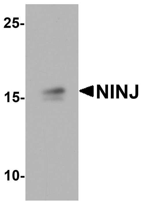
| Calculated MW | 16345 Da |
| Concentration (mg/ml) | 1 mg/mL |
| Conjugate | Unconjugated |
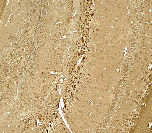
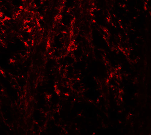
| Application Notes | NINJ1 antibody can be used for detection of NINJ1 by Western blot at 1 - 2 µg/ml. Antibody can also be used for immunohistochemistry starting at 5 µg/mL. For immunofluorescence start at 20 µg/mL. |
| Gene ID | 4814 |
|---|---|
| Other Names | Ninjurin-1, Nerve injury-induced protein 1, NINJ1 |
| Target/Specificity | NINJ1; NINJ1 antibody is human, mouse and rat reactive. |
| Reconstitution & Storage | NINJ1 antibody can be stored at 4℃ for three months and -20℃, stable for up to one year. |
| Precautions | NINJ1 Antibody is for research use only and not for use in diagnostic or therapeutic procedures. |
For Research Use Only. Not For Use In Diagnostic Procedures.
| Name | NINJ1 {ECO:0000303|PubMed:33472215, ECO:0000312|HGNC:HGNC:7824} |
|---|---|
| Function | [Ninjurin-1]: Effector of various programmed cell death, such as pyroptosis and necroptosis, which mediates plasma membrane rupture (cytolysis) (PubMed:33472215, PubMed:36468682, PubMed:37196676, PubMed:37198476, PubMed:38614101, PubMed:39667936). Oligomerizes in response to death stimuli and forms ring-like structures on the plasma membrane: acts by cutting and shedding membrane disks, like a cookie cutter, leading to membrane damage and loss that cannot be repaired by the cell (PubMed:38614101). Plasma membrane rupture leads to release intracellular molecules named damage-associated molecular patterns (DAMPs) that propagate the inflammatory response (PubMed:33472215, PubMed:36468682, PubMed:37196676, PubMed:37198476). Mechanistically, mediates plasma membrane rupture by introducing hydrophilic faces of 2 alpha helices into the hydrophobic membrane (PubMed:37198476, PubMed:38614101). Induces plasma membrane rupture downstream of Gasdermin (GSDMA, GSDMB, GSDMC, GSDMD, or GSDME) or MLKL during pyroptosis or necroptosis, respectively (PubMed:33472215, PubMed:36468682, PubMed:37196676, PubMed:37198476). Acts as an effector of PANoptosis downstream of CASP1, CASP4, CASP8 and RIPK3 (By similarity). Also induces plasma membrane rupture in response to cell swelling caused by osmotic stress and ferroptosis downstream of lipid peroxidation (By similarity). Acts as a regulator of Toll-like receptor 4 (TLR4) signaling triggered by lipopolysaccharide (LPS) during systemic inflammation; directly binds LPS (PubMed:26677008). Involved in leukocyte migration during inflammation by promoting transendothelial migration of macrophages via homotypic binding (By similarity). Promotes the migration of monocytes across the brain endothelium to central nervous system inflammatory lesions (PubMed:22162058). Also acts as a homophilic transmembrane adhesion molecule involved in various processes such as axonal growth, cell chemotaxis and angiogenesis (PubMed:33028854, PubMed:8780658, PubMed:9261151). Promotes cell adhesion by mediating homophilic interactions via its extracellular N-terminal adhesion motif (N-NAM) (PubMed:33028854, PubMed:8780658, PubMed:9261151). Involved in the progression of the inflammatory stress by promoting cell-to-cell interactions between immune cells and endothelial cells (PubMed:22162058, PubMed:26677008, PubMed:32147432). Plays a role in nerve regeneration by promoting maturation of Schwann cells (PubMed:8780658, PubMed:9261151). Acts as a regulator of angiogenesis (PubMed:33028854). Promotes the formation of new vessels by mediating the interaction between capillary pericyte cells and endothelial cells (By similarity). Promotes osteoclasts development by enhancing the survival of prefusion osteoclasts (By similarity). Also involved in striated muscle growth and differentiation (By similarity). |
| Cellular Location | [Ninjurin-1]: Cell membrane; Multi-pass membrane protein. Synaptic cell membrane {ECO:0000250|UniProtKB:O70131}; Multi-pass membrane protein |
| Tissue Location | Widely expressed in both adult and embryonic tissues, primarily those of epithelial origin |
Provided below are standard protocols that you may find useful for product applications.
BACKGROUND
Ninjurin-1 (NINJ1) is a homophilic adhesion molecule that is strongly induced in dorsal root ganglion (DRG) and Schwann cells following nerve injury and promotes neurite outgrowth of DRG neurons (1,2), suggesting that NINJ1 may play a role in nerve regeneration. NINJ1 is transcriptionally regulated by p53 and is induced by DNA damage in a p53-dependent manner. A deficiency of NINJ1 increases p53 expression, suppresses cell proliferation, but enhances apoptosis and premature senescence in a p53-dependent manner as well as radiation-induced cell mortality (3).
REFERENCES
Araki T and Milbrandt J. Ninjurin, a novel adhesion molecule, is induced by nerve injury and promotes axonal growth. Neuron 1996; 17:353-61.
Araki T, Zimonjic DB, Popescu NC, et al. Mechanism of homophilic binding mediated by ninjurin, a novel widely expressed adhesion molecule. J. Biol. Chem. 1997; 212:21373-80.
Cho SJ, Rossi A, Jung YS, et al. Ninjurin1, a target of p53, regulates p53 expression and p53-dependent cell survival, sensecence, and radiation-induced mortality. Proc. Natl. Acad. Sci. USA 2013; 110:9362-7.

终于等到您。ABCEPTA(百远生物)抗体产品。
点击下方“我要评价 ”按钮提交您的反馈信息,您的反馈和评价是我们最宝贵的财富之一,
我们将在1-3个工作日内处理您的反馈信息。
如有疑问,联系:0512-88856768 tech-china@abcepta.com.


