


 癌症的基本特征包括细胞增殖、血管生成、迁移、凋亡逃避机制和细胞永生等。找到癌症发生过程中这些通路的关键标记物和对应的抗体用于检测至关重要。
 为您推荐一个泛素化位点预测神器——泛素化分析工具,可以为您的蛋白的泛素化位点作出预测和评分。
 细胞自噬受体图形绘图工具为你的蛋白的细胞受体结合位点作出预测和评分,识别结合到自噬通路中的蛋白是非常重要的,便于让我们理解自噬在正常生理、病理过程中的作用,如发育、细胞分化、神经退化性疾病、压力条件下、感染和癌症。
KIRREL3 Antibody


- 产品详情
- 实验流程
- 背景知识
Application
| WB, IF, E, IHC-P |
|---|---|
| Primary Accession | Q8IZU9 |
| Other Accession | NP_115920, 26006461 |
| Reactivity | Human, Mouse, Rat |
| Host | Rabbit |
| Clonality | Polyclonal |
| Isotype | IgG |
| Calculated MW | 85255 Da |
| Concentration (mg/ml) | 1 mg/mL |
| Conjugate | Unconjugated |
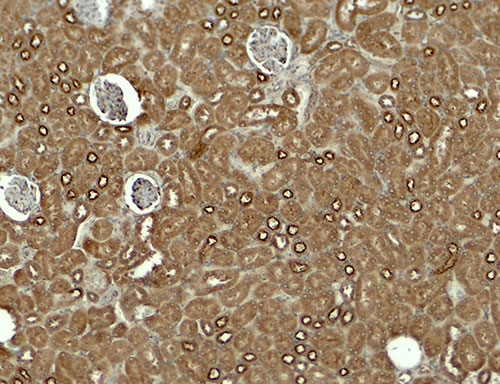
| Application Notes | KIRREL3 antibody can be used for detection of KIRREL3 by Western blot at 1 - 2 µg/ml. Antibody can also be used for immunohistochemistry starting at 5 µg/mL. For immunofluorescence start at 20 µg/mL. |
| Gene ID | 84623 |
|---|---|
| Other Names | Kin of IRRE-like protein 3, Kin of irregular chiasm-like protein 3, Nephrin-like protein 2, KIRREL3, KIAA1867, NEPH2 |
| Target/Specificity | KIRREL3; KIRREL3 antibody is human, mouse and rat reactive. At least two isoforms are known to exist; this antibody will detect both isoforms. KIRREL3 antibody is predicted to not cross-react with other members of the KIRREL protein family. |
| Reconstitution & Storage | KIRREL3 antibody can be stored at 4℃ for three months and -20℃, stable for up to one year. |
| Precautions | KIRREL3 Antibody is for research use only and not for use in diagnostic or therapeutic procedures. |
| Name | KIRREL3 (HGNC:23204) |
|---|---|
| Function | Synaptic adhesion molecule required for the formation of target-specific synapses. Required for formation of target-specific synapses at hippocampal mossy fiber synapses. Required for formation of mossy fiber filopodia, the synaptic structures connecting dentate granule and GABA neurons. Probably acts as a homophilic adhesion molecule that promotes trans-cellular interactions and stabilize mossy fiber filipodia contact and subsequent synapse formation. Required for the coalescence of vomeronasal sensory neuron axons. May be involved in the hematopoietic supportive capacity of stroma cells; the secreted extracellular domain is directly responsible for supporting hematopoietic stem cells. |
| Cellular Location | Cell membrane; Single-pass type I membrane protein |
| Tissue Location | Expressed in fetal and adult brain (PubMed:19012874). Also expressed in kidney, specifically in podocytes of kidney glomeruli (PubMed:12424224). Also expressed in skeletal muscle (PubMed:25488023). |
For Research Use Only. Not For Use In Diagnostic Procedures.
Provided below are standard protocols that you may find useful for product applications.
BACKGROUND
KIRREL3, also known as nephrin-like protein 2, is a type I transmembrane protein belonging to a family of three podocin interacting proteins and the immunoglobulin superfamily (1). KIRREL3 is involved in the regulation of both glomerular and neural development (2), and more specifically, the nucleogenesis of the pontine nuclei in the developing hindbrain (3). KIRREL3 has also been shown to interact with the synaptic scaffold protein calmodulin-associated serine/threonine kinase (CASK) in neuronal cells (4).
REFERENCES
Sellin L, Huber TB, Gerke P, et al. NEPH1 defines a novel family of podocin interacting proteins. FASEB J. 2003; 17:115-7.
Neumann-Haefelin E, Kramer-Zucker A, Slanchev K, et al. A model organism approach: defining the role of Neph proteins as regulators of neuron and kidney morphogenesis. Hum. Mol. Genet. 2010; 19:2347-59.
Nishida K, Nakayama K, Yoshimura S, et al. Role of Neph2 in pontine nuclei formation in the developing hindbrain. Mol. Cell Neurosci. 2011; 46:662-70.
Mizuhara E, Minaki Y, Nakatani T, et al. Purkinje cells originate from cerebellar ventricular zone progenitors positive for Neph3 and E-cadherin. Dev. Biol. 2010; 338:202-14.
终于等到您。ABCEPTA(百远生物)抗体产品。
点击下方“我要评价 ”按钮提交您的反馈信息,您的反馈和评价是我们最宝贵的财富之一,
我们将在1-3个工作日内处理您的反馈信息。
如有疑问,联系:0512-88856768 tech-china@abcepta.com.


