


 癌症的基本特征包括细胞增殖、血管生成、迁移、凋亡逃避机制和细胞永生等。找到癌症发生过程中这些通路的关键标记物和对应的抗体用于检测至关重要。
 为您推荐一个泛素化位点预测神器——泛素化分析工具,可以为您的蛋白的泛素化位点作出预测和评分。
 细胞自噬受体图形绘图工具为你的蛋白的细胞受体结合位点作出预测和评分,识别结合到自噬通路中的蛋白是非常重要的,便于让我们理解自噬在正常生理、病理过程中的作用,如发育、细胞分化、神经退化性疾病、压力条件下、感染和癌症。
EDA1 Antibody
- 产品详情
- 实验流程
- 背景知识
Application
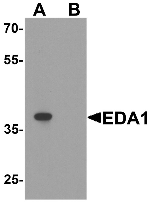
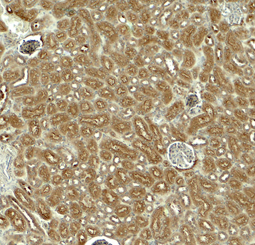
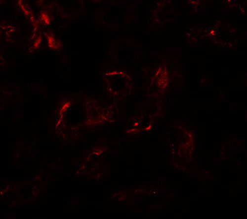
| WB, IF, E, IHC-P |
|---|---|
| Primary Accession | Q92838 |
| Other Accession | NP_001390, 4503449 |
| Reactivity | Human, Mouse, Rat |
| Host | Rabbit |
| Clonality | Polyclonal |
| Isotype | IgG |
| Calculated MW | 41294 Da |
| Concentration (mg/ml) | 1 mg/mL |
| Conjugate | Unconjugated |
| Application Notes | EDA1 antibody can be used for detection of EDA1 by Western blot at 1 - 2 µg/ml. Antibody can also be used for immunohistochemistry starting at 5 µg/mL. For immunofluorescence start at 20 µg/mL. |
| Gene ID | 1896 |
|---|---|
| Other Names | Ectodysplasin-A, Ectodermal dysplasia protein, EDA protein, Ectodysplasin-A, membrane form, Ectodysplasin-A, secreted form, EDA, ED1, EDA2 |
| Target/Specificity | EDA; EDA1 antibody is human, mouse and rat reactive. Multiple isoforms of EDA1 are known to exist. |
| Reconstitution & Storage | EDA1 antibody can be stored at 4℃ for three months and -20℃, stable for up to one year. |
| Precautions | EDA1 Antibody is for research use only and not for use in diagnostic or therapeutic procedures. |
| Name | EDA |
|---|---|
| Synonyms | ED1, EDA2 |
| Function | Cytokine which is involved in epithelial-mesenchymal signaling during morphogenesis of ectodermal organs. Functions as a ligand activating the DEATH-domain containing receptors EDAR and EDA2R (PubMed:11039935, PubMed:27144394, PubMed:34582123, PubMed:8696334). May also play a role in cell adhesion (By similarity). |
| Cellular Location | Cell membrane {ECO:0000250|UniProtKB:O54693}; Single-pass type II membrane protein {ECO:0000250|UniProtKB:O54693} |
| Tissue Location | Not abundant; expressed in specific cell types of ectodermal (but not mesodermal) origin of keratinocytes, hair follicles, sweat glands. Also in adult heart, liver, muscle, pancreas, prostate, fetal liver, uterus, small intestine and umbilical cord {ECO:0000269|Ref.6} |
For Research Use Only. Not For Use In Diagnostic Procedures.
Provided below are standard protocols that you may find useful for product applications.
BACKGROUND
Ectodysplasin A (EDA1) is a member of the TNF-related ligand family involved in the early epithelial-mesenchymal interaction that regulates ectodermal appendage formation (1). It is a trimeric type II membrane protein that co-localizes with cytoskeletal structures at the lateral and apical surfaces of cells and can be expressed as eight alternatively spliced isoforms in hair follicles and in the epidermis of adult skin (2,3). EDAs are required during development, and loss or mutation of EDA1 results in a group of developmental disorders identified as ectodermal dysplasia type 1 (4,5).
REFERENCES
Kere J, Srivastava AK, Montonen O. X-linked anhidrotic (hypohidrotic) ectodermal dysplasia is caused by mutation in a novel transmembrane protein. Nat. Genet. 1996; 13:409-16.
Vincent MC, Biancalana V, Ginisty D, et al. Mutational spectrum of the ED1 gene in X-linked hypohidrotic ectodermal dysplasia. Eur. J. Hum. Genet. 2001; 9:355-63.
Ohashi M, Moriya C, Tanahashi K, et al. A new EDA gene mutation in a family of X-linked hypohidrotic ectodermal dysplasia. J. Dermatol. Sci. 2014; 74:175-7.
Bayés M, Hartung AJ, Ezer S, et al. The anhidrotic ectodermal dysplasia gene (EDA) undergoes alternative splicing and encodes ectodysplasin-A with deletion mutations in collagenous repeats. Hum. Mol. Genet. 1998; 7:1661-9.
终于等到您。ABCEPTA(百远生物)抗体产品。
点击下方“我要评价 ”按钮提交您的反馈信息,您的反馈和评价是我们最宝贵的财富之一,
我们将在1-3个工作日内处理您的反馈信息。
如有疑问,联系:0512-88856768 tech-china@abcepta.com.


