


 癌症的基本特征包括细胞增殖、血管生成、迁移、凋亡逃避机制和细胞永生等。找到癌症发生过程中这些通路的关键标记物和对应的抗体用于检测至关重要。
 为您推荐一个泛素化位点预测神器——泛素化分析工具,可以为您的蛋白的泛素化位点作出预测和评分。
 细胞自噬受体图形绘图工具为你的蛋白的细胞受体结合位点作出预测和评分,识别结合到自噬通路中的蛋白是非常重要的,便于让我们理解自噬在正常生理、病理过程中的作用,如发育、细胞分化、神经退化性疾病、压力条件下、感染和癌症。
Hexokinase 1 Antibody
- 产品详情
- 实验流程
- 背景知识
Application
| WB, IHC, IF, E |
|---|---|
| Primary Accession | P19367 |
| Other Accession | NP_000179, 188497754 |
| Reactivity | Human, Mouse, Rat |
| Host | Rabbit |
| Clonality | Polyclonal |
| Isotype | IgG |
| Calculated MW | 102486 Da |
| Concentration (mg/ml) | 1 mg/mL |
| Conjugate | Unconjugated |
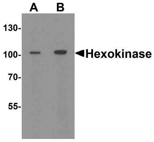
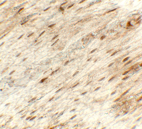
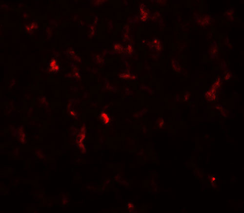
| Application Notes | Hexokinase 1 antibody can be used for detection of Hexokinase 1 by Western blot at 1 - 2 µg/ml. Antibody can also be used for immunohistochemistry starting at 5 µg/mL. For immunofluorescence start at 20 µg/mL. |
| Gene ID | 3098 |
|---|---|
| Other Names | Hexokinase-1, 2.7.1.1, Brain form hexokinase, Hexokinase type I, HK I, HK1 |
| Target/Specificity | HK1; Hexokinase 1 antibody is human, mouse and rat reactive. Multiple isoforms of Hexokinase 1 are known to exist. |
| Reconstitution & Storage | Hexokinase 1 antibody can be stored at 4℃ for three months and -20℃, stable for up to one year. |
| Precautions | Hexokinase 1 Antibody is for research use only and not for use in diagnostic or therapeutic procedures. |
| Name | HK1 (HGNC:4922) |
|---|---|
| Function | Catalyzes the phosphorylation of various hexoses, such as D- glucose, D-glucosamine, D-fructose, D-mannose and 2-deoxy-D-glucose, to hexose 6-phosphate (D-glucose 6-phosphate, D-glucosamine 6-phosphate, D-fructose 6-phosphate, D-mannose 6-phosphate and 2-deoxy-D-glucose 6- phosphate, respectively) (PubMed:1637300, PubMed:25316723, PubMed:27374331). Does not phosphorylate N-acetyl-D-glucosamine (PubMed:27374331). Mediates the initial step of glycolysis by catalyzing phosphorylation of D-glucose to D-glucose 6-phosphate (By similarity). Involved in innate immunity and inflammation by acting as a pattern recognition receptor for bacterial peptidoglycan (PubMed:27374331). When released in the cytosol, N-acetyl-D-glucosamine component of bacterial peptidoglycan inhibits the hexokinase activity of HK1 and causes its dissociation from mitochondrial outer membrane, thereby activating the NLRP3 inflammasome (PubMed:27374331). |
| Cellular Location | Mitochondrion outer membrane; Peripheral membrane protein. Cytoplasm, cytosol. Note=The mitochondrial-binding peptide (MBP) region promotes association with the mitochondrial outer membrane (Probable). Dissociates from the mitochondrial outer membrane following inhibition by N-acetyl-D-glucosamine, leading to relocation to the cytosol (PubMed:27374331). |
| Tissue Location | Isoform 2: Erythrocyte specific (Ref.6). Isoform 3: Testis-specific (PubMed:10978502). Isoform 4: Testis-specific (PubMed:10978502). {ECO:0000269|PubMed:10978502, ECO:0000269|Ref.6} |
For Research Use Only. Not For Use In Diagnostic Procedures.
Provided below are standard protocols that you may find useful for product applications.
BACKGROUND
There are four major glucose-phosphorylating isoenzymes, designated Hexokinase 1 I, II, III, and IV (1). Hexokinase 1 activity is involved in the first step in several metabolic pathways including phosphorylation of glucose to produce glucose-6-phosphate, thus committing glucose to the glycolytic pathway (1,2). Hexokinase 1 2 is the predominant Hexokinase 1 isozyme expressed in insulin-responsive tissues such as skeletal muscle and its expression is insulin-responsive (3). It is involved in the increased rate of glycolysis seen in rapidly growing cancer cells (4).
REFERENCES
Chen T, Ning D, Sun H, et al. Sequence Analysis and Molecular Characterization of Clonorchis sinensis Hexokinase 1, an Unusual Trimeric 50-kDa Glucose-6-Phosphate-Sensitive Allosteric Enzyme. PLoS One 2014; 9:e107940.
Halestrap AP, Pereira GC, and Pasdois P. The role of Hexokinase 1 in cardioprotection-mechanism and potential for translation. Br. J. Pharmacol. 2014; epub.
Roberts DJ and Miyamoto S. Hexokinase 1 II integrates energy metabolism and cellular protection: Akting on mitochondria and TORCing to autophagy. Cell Death Differ. 2014; epub.
Wang L, Xiong H, Wu F, et al. Hexokinase 1 2-Mediated Warburg Effect Is Required for PTEN- and p53-Deficiency-Driven Prostate Cancer Growth. Cell Rep. 2014; 8:1461-74.
终于等到您。ABCEPTA(百远生物)抗体产品。
点击下方“我要评价 ”按钮提交您的反馈信息,您的反馈和评价是我们最宝贵的财富之一,
我们将在1-3个工作日内处理您的反馈信息。
如有疑问,联系:0512-88856768 tech-china@abcepta.com.


