


 癌症的基本特征包括细胞增殖、血管生成、迁移、凋亡逃避机制和细胞永生等。找到癌症发生过程中这些通路的关键标记物和对应的抗体用于检测至关重要。
 为您推荐一个泛素化位点预测神器——泛素化分析工具,可以为您的蛋白的泛素化位点作出预测和评分。
 细胞自噬受体图形绘图工具为你的蛋白的细胞受体结合位点作出预测和评分,识别结合到自噬通路中的蛋白是非常重要的,便于让我们理解自噬在正常生理、病理过程中的作用,如发育、细胞分化、神经退化性疾病、压力条件下、感染和癌症。
VTI1b Antibody
- 产品详情
- 实验流程
- 背景知识
Application
| WB, IHC, E |
|---|---|
| Primary Accession | Q9UEU0 |
| Other Accession | NP_006361, 5454166 |
| Reactivity | Human, Mouse, Rat |
| Host | Rabbit |
| Clonality | Polyclonal |
| Isotype | IgG |
| Calculated MW | 26688 Da |
| Concentration (mg/ml) | 1 mg/mL |
| Conjugate | Unconjugated |
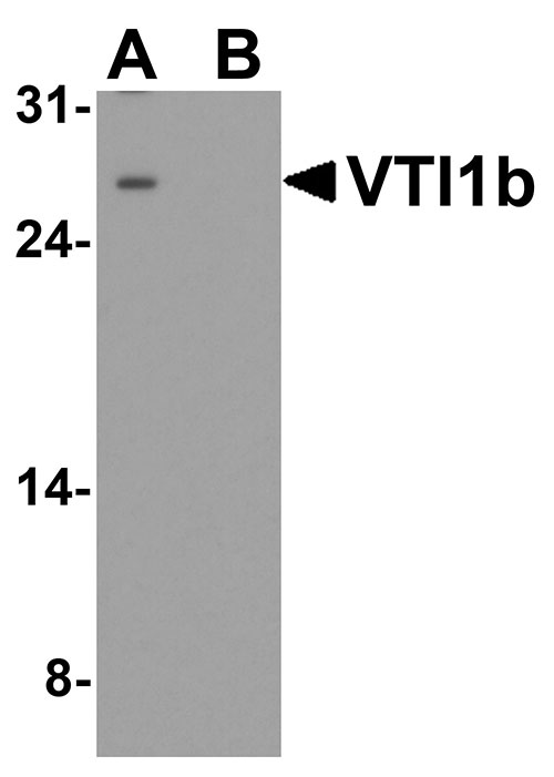
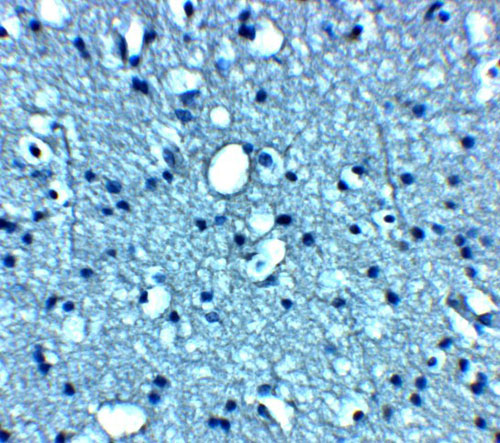
| Application Notes | VTI1b antibody can be used for detection of VTI1b by Western blot at 1 - 2 µg/ml. Antibody can also be used for immunohistochemistry starting at 5 µg/mL. |
| Gene ID | 10490 |
|---|---|
| Other Names | Vesicle transport through interaction with t-SNAREs homolog 1B, Vesicle transport v-SNARE protein Vti1-like 1, Vti1-rp1, VTI1B, VTI1, VTI1L, VTI1L1, VTI2 |
| Target/Specificity | VTI1b; VTI1b antibody is human, mouse and rat reactive. At least three isoforms of VTI1a are known to exist; this antibody will detect all three isoforms. VTI1b antibody is predicted to not cross-react with VTI1a. |
| Reconstitution & Storage | VTI1b antibody can be stored at 4℃ for three months and -20℃, stable for up to one year. |
| Precautions | VTI1b Antibody is for research use only and not for use in diagnostic or therapeutic procedures. |
| Name | VTI1B |
|---|---|
| Synonyms | VTI1, VTI1L, VTI1L1, VTI2 |
| Function | V-SNARE that mediates vesicle transport pathways through interactions with t-SNAREs on the target membrane. These interactions are proposed to mediate aspects of the specificity of vesicle trafficking and to promote fusion of the lipid bilayers. May be concerned with increased secretion of cytokines associated with cellular senescence. |
| Cellular Location | Early endosome membrane; Single-pass type IV membrane protein. Late endosome membrane; Single-pass type IV membrane protein. Lysosome membrane. Cytoplasmic granule. Recycling endosome membrane; Single-pass type IV membrane protein |
| Tissue Location | Expressed in all tissues examined. |
For Research Use Only. Not For Use In Diagnostic Procedures.
Provided below are standard protocols that you may find useful for product applications.
BACKGROUND
Vesicle transport through interaction with t-SNAREs homolog 1 (VTI1a and VTI1b) are involved in vesicular transport from the late endosomes to the trans-Golgi network (1). They are both localized in the trans-Golgi network, with VTI1a also found in the Golgi apparatus and VTI1b in endosomes (2,3). It is thought that VTI1b along with the SNARE protein VAMP8 mediates the fusion of antimicrobial and canonical autophagosomes with lysosomes, an essential process for autophagy (4).
REFERENCES
Fischer VM and Stevens TH. A human homolog can functionally replace the yeast vesicle-associated SNARE Vti1p in two vesicle transport pathways. J. Biol. Chem. 1998; 273:2624-30.
Kreykenbohm V, Wenzel D, Antonin W et al. The SNAREs vti1a and vti1b have distinct localization and SNARE complex partners. Eur. J. Cell Biol. 2002; 81:273-80.
Antonin W, Riedel D, von Mollard GF, et al. The SNARE Vti1a-beta is localized to small synaptic vesicles and participates in a novel SNARE complex. J. Neurosci. 2000; 20:5724-32.
Furuta N, Fujita N, Noda T, et al. Combinational soluble N-ethylmaleimide-sensitive factor attachment protein receptor proteins VAMP8 and Vti1b mediate fusion of antimicrobial and canonical autophagosomes with lysosomes. Mol. Biol. Cell. 2010; 21:1001-10.
终于等到您。ABCEPTA(百远生物)抗体产品。
点击下方“我要评价 ”按钮提交您的反馈信息,您的反馈和评价是我们最宝贵的财富之一,
我们将在1-3个工作日内处理您的反馈信息。
如有疑问,联系:0512-88856768 tech-china@abcepta.com.


