


 癌症的基本特征包括细胞增殖、血管生成、迁移、凋亡逃避机制和细胞永生等。找到癌症发生过程中这些通路的关键标记物和对应的抗体用于检测至关重要。
 为您推荐一个泛素化位点预测神器——泛素化分析工具,可以为您的蛋白的泛素化位点作出预测和评分。
 细胞自噬受体图形绘图工具为你的蛋白的细胞受体结合位点作出预测和评分,识别结合到自噬通路中的蛋白是非常重要的,便于让我们理解自噬在正常生理、病理过程中的作用,如发育、细胞分化、神经退化性疾病、压力条件下、感染和癌症。
TMPRSS2 (CT) Antibody
Infectious Disease, COVID-19

- 产品详情
- 实验流程
- 背景知识
Application
| WB, IF, E |
|---|---|
| Primary Accession | O15393 |
| Other Accession | O15393 |
| Reactivity | Rat |
| Host | Rabbit |
| Clonality | Polyclonal |
| Isotype | IgG |
| Clone Names | TMPRSS2 |
| Calculated MW | 53859 Da |
| Concentration (mg/ml) | 1 mg/mL |
| Conjugate | Unconjugated |
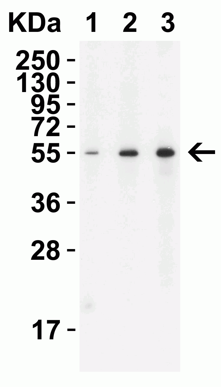
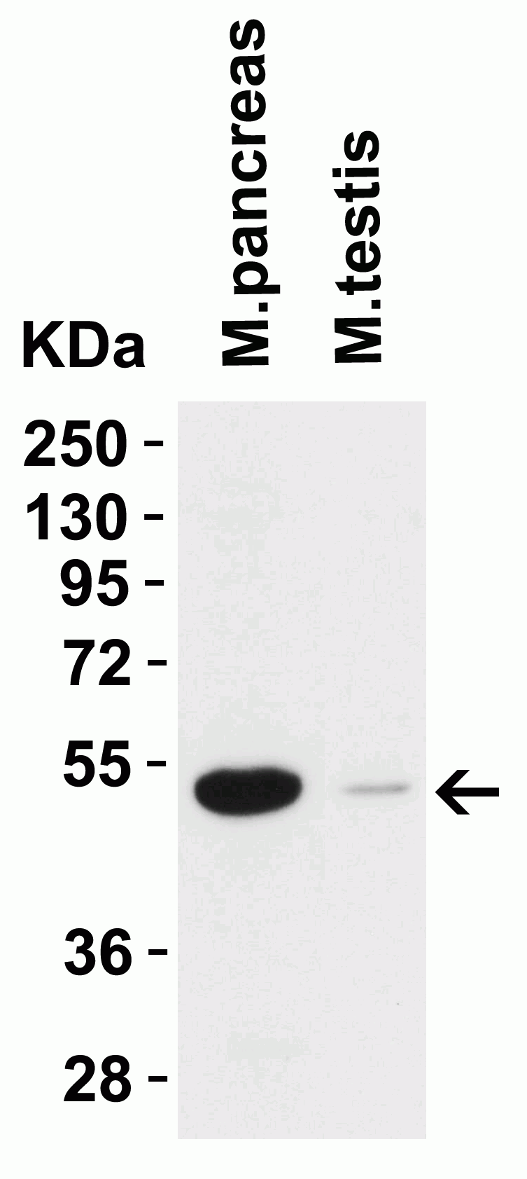
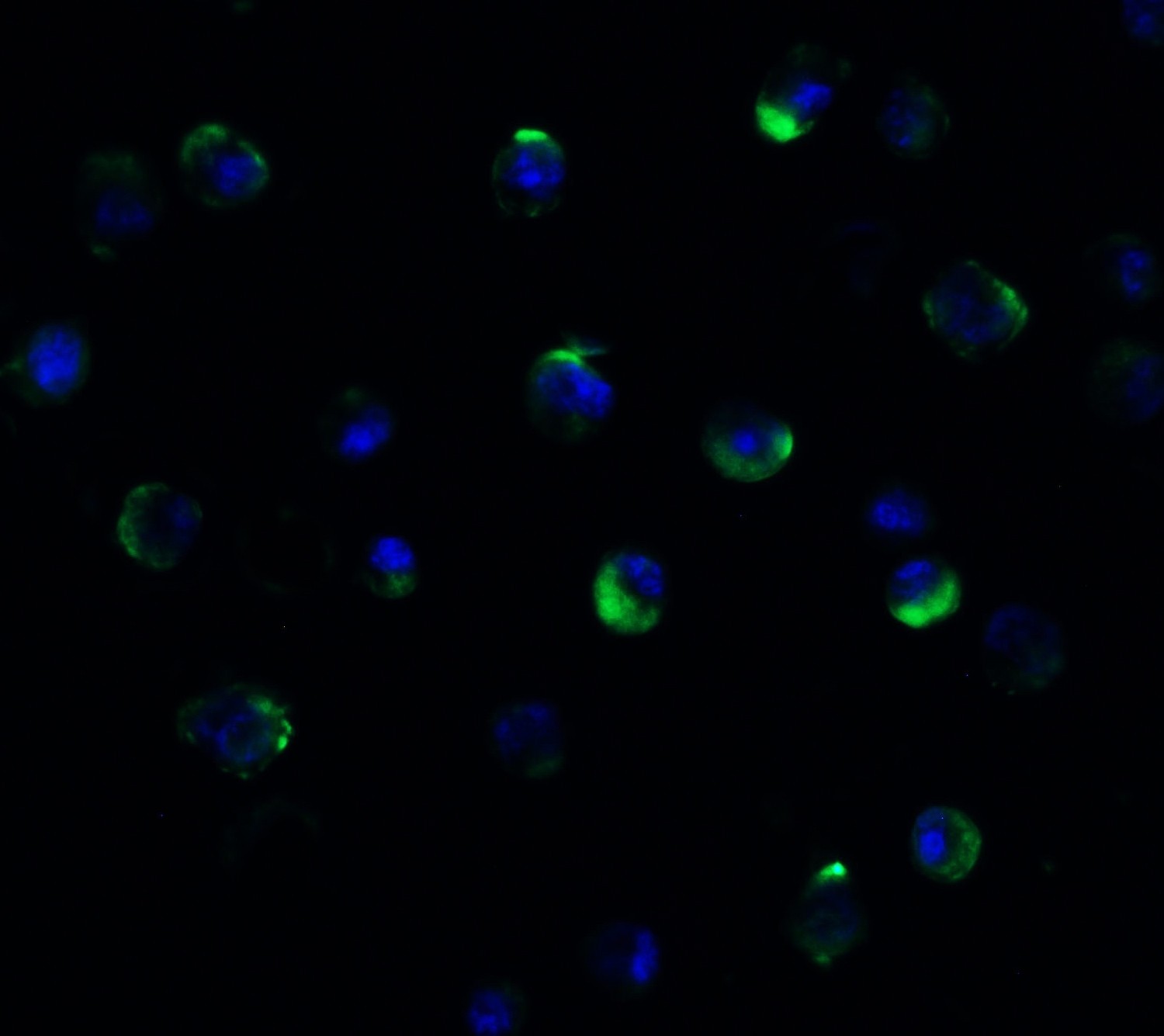
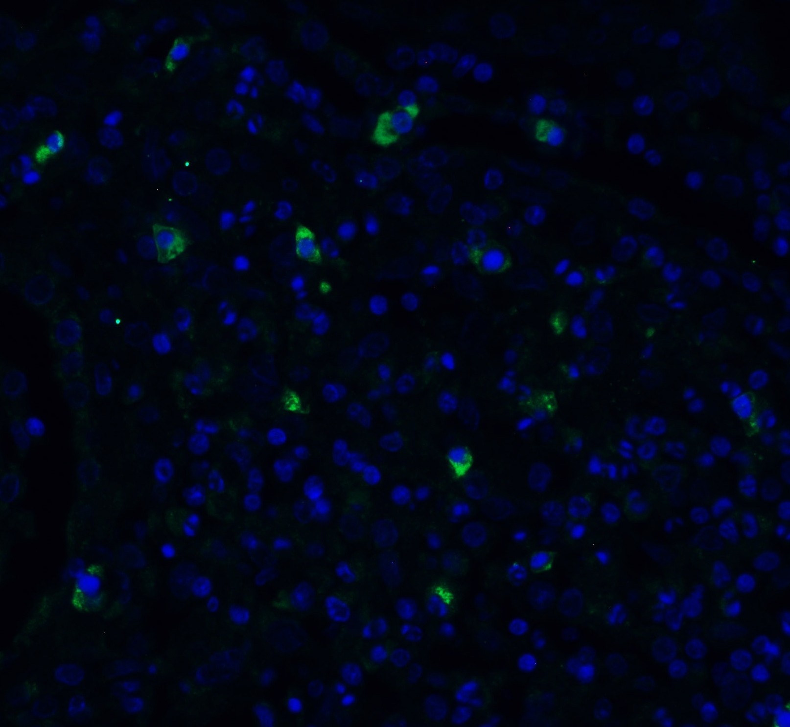
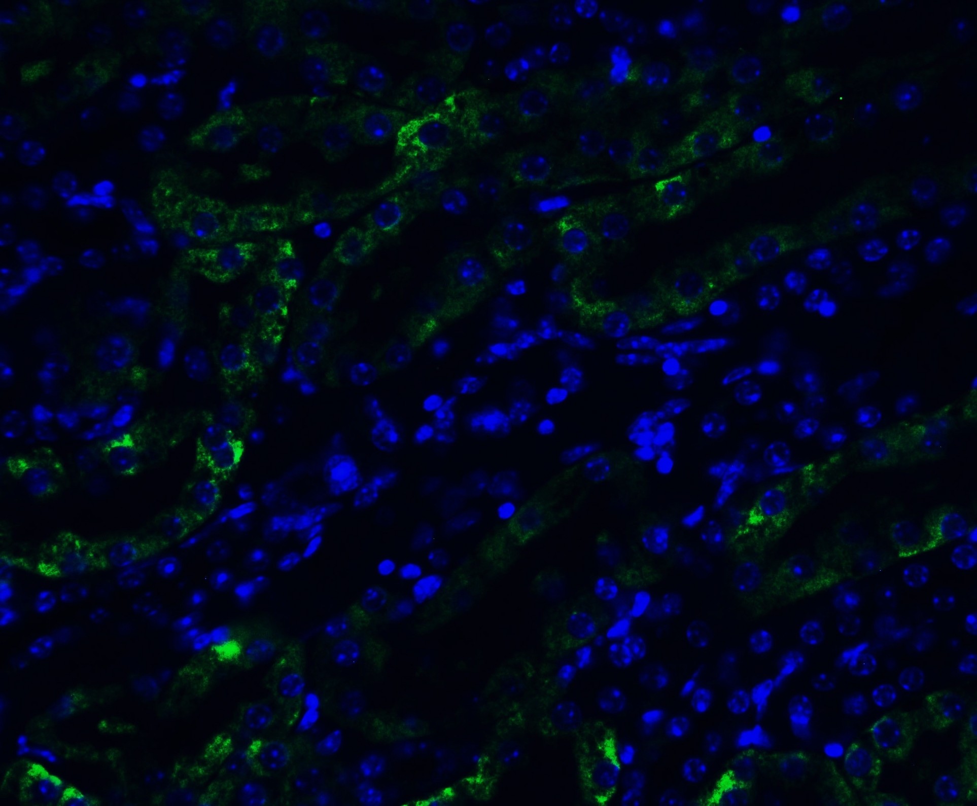
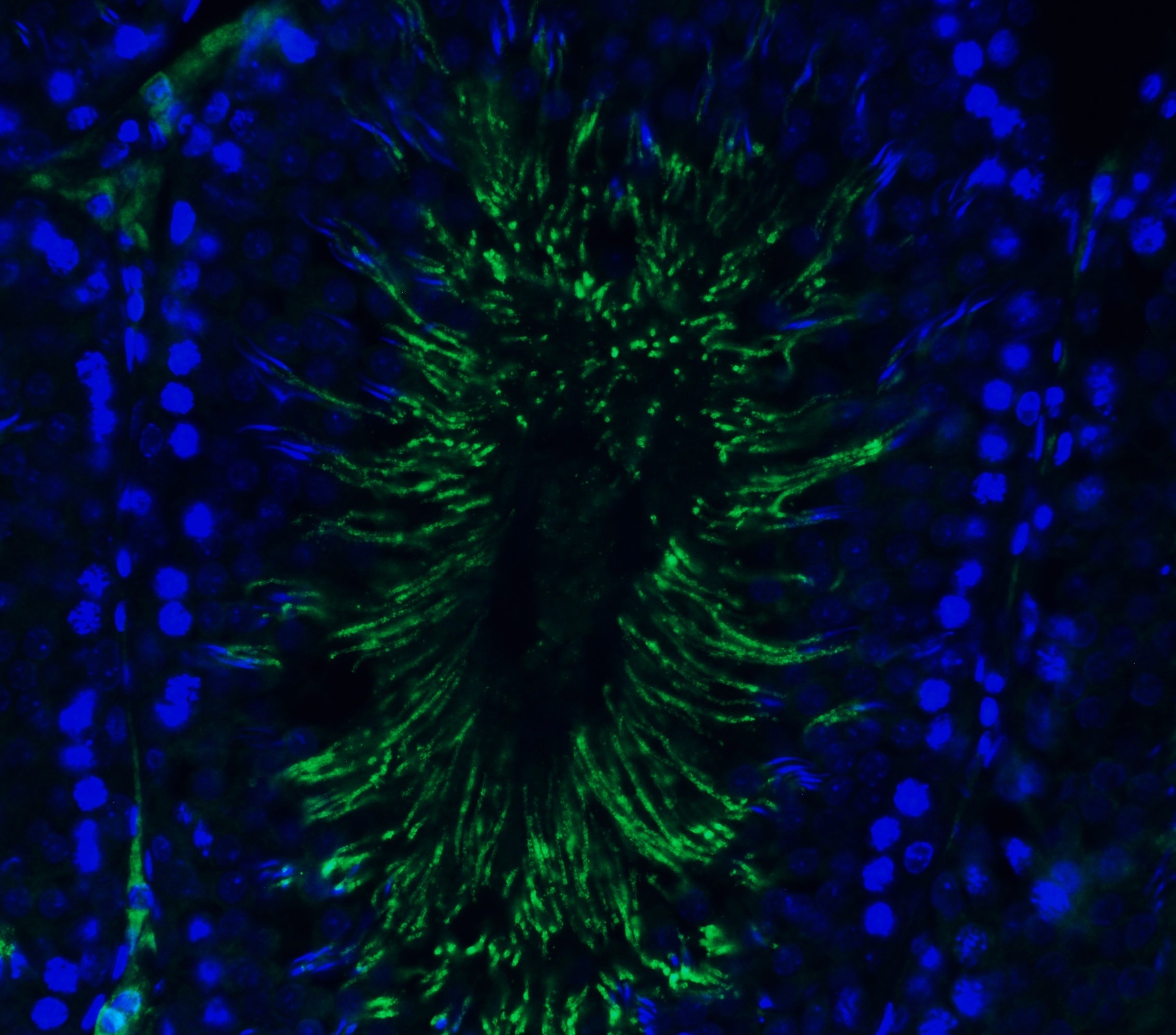
| Application Notes | WB: 2 μg/mL; IF: 20 μg/mL. Antibody validated: Western Blot in human, mouse and rat samples; Immunofluorescence in human, mouse and rat samples. All other applications and species not yet tested. |
| Gene ID | 7113 |
|---|---|
| Alias Symbol | TMPRSS2 |
| Other Names | TMPRSS2 Antibody: Transmembrane protease serine 2, Serine protease 10, PRSS10, Transmembrane protease serine 2 non-catalytic chain, Transmembrane protease serine 2 catalytic chain. |
| Reconstitution & Storage | TMPRSS2 antibody can be stored at 4˚C for three months and -20˚C, stable for up to one year. As with all antibodies care should be taken to avoid repeated freeze thaw cycles. Antibodies should not be exposed to prolonged high temperatures. |
| Precautions | TMPRSS2 (CT) Antibody is for research use only and not for use in diagnostic or therapeutic procedures. |
| Name | TMPRSS2 (HGNC:11876) |
|---|---|
| Synonyms | PRSS10 |
| Function | Plasma membrane-anchored serine protease that cleaves at arginine residues (PubMed:32703818, PubMed:35676539, PubMed:37990007, PubMed:38964328). Participates in proteolytic cascades of relevance for the normal physiologic function of the prostate (PubMed:25122198). Androgen-induced TMPRSS2 activates several substrates that include pro- hepatocyte growth factor/HGF, the protease activated receptor-2/F2RL1 or matriptase/ST14 leading to extracellular matrix disruption and metastasis of prostate cancer cells (PubMed:15537383, PubMed:25122198, PubMed:26018085). In addition, activates trigeminal neurons and contribute to both spontaneous pain and mechanical allodynia (By similarity). |
| Cellular Location | Cell membrane; Single-pass type II membrane protein |
| Tissue Location | Expressed in several tissues that comprise large populations of epithelial cells with the highest level of transcripts measured in the prostate gland. Expressed in type II pneumocytes in the lung (at protein level). Expressed strongly in small intestine. Also expressed in colon, stomach and salivary gland. Coexpressed with ACE2 within lung type II pneumocytes, ileal absorptive enterocytes, intestinal epithelial cells, cornea, gallbladder and nasal goblet secretory cells (Ref.21). {ECO:0000269|PubMed:11169526, ECO:0000269|PubMed:20382709, ECO:0000269|PubMed:21325420, ECO:0000269|PubMed:32404436, ECO:0000269|Ref.21} |
For Research Use Only. Not For Use In Diagnostic Procedures.
Provided below are standard protocols that you may find useful for product applications.
BACKGROUND
TMPRSS2 Antibody: TMPRSS2 is a plasma membrane-anchored serine protease that participates in proteolytic cascades of relevance for the normal physiologic function of the prostate. Androgen-induced TMPRSS2 activates several substrates that include pro-hepatocyte growth factor/HGF, the protease activated receptor-2/F2RL1 or matriptase/ST14 leading to extracellular matrix disruption and metastasis of prostate cancer cells. It facilitates human coronaviruses SARS-CoV and SARS-CoV-2 infections via two independent mechanisms, proteolytic cleavage of ACE2 receptor which promotes viral uptake, and cleavage of coronavirus spike glycoproteins which activates the glycoprotein for host cell entry. It proteolytically cleaves and activates the spike glycoproteins of human coronavirus 229E (HCoV-229E) and human coronavirus EMC (HCoV-EMC) and the fusion glycoproteins F0 of Sendai virus (SeV), human metapneumovirus (HMPV), human parainfluenza 1, 2, 3, 4a and 4b viruses (HPIV). TMPRSS2 is essential for spread and pathogenesis of influenza A virus (strains H1N1, H3N2 and H7N9), and it is involved in proteolytic cleavage and activation of hemagglutinin (HA) protein which is essential for viral infectivity.
REFERENCES
Lucas et al. Cancer Discov. 2014; 4(11):1310-25.
Ko et al. Cancer Res. 2015; 75(14):2949-60.
Zang et al. Sci. Immunol. 2020; 5(47):eabc3582.
终于等到您。ABCEPTA(百远生物)抗体产品。
点击下方“我要评价 ”按钮提交您的反馈信息,您的反馈和评价是我们最宝贵的财富之一,
我们将在1-3个工作日内处理您的反馈信息。
如有疑问,联系:0512-88856768 tech-china@abcepta.com.


