


 癌症的基本特征包括细胞增殖、血管生成、迁移、凋亡逃避机制和细胞永生等。找到癌症发生过程中这些通路的关键标记物和对应的抗体用于检测至关重要。
 为您推荐一个泛素化位点预测神器——泛素化分析工具,可以为您的蛋白的泛素化位点作出预测和评分。
 细胞自噬受体图形绘图工具为你的蛋白的细胞受体结合位点作出预测和评分,识别结合到自噬通路中的蛋白是非常重要的,便于让我们理解自噬在正常生理、病理过程中的作用,如发育、细胞分化、神经退化性疾病、压力条件下、感染和癌症。
SARS-CoV-2 (COVID-19) ORF3a Antibody
Infectious Disease, COVID-19
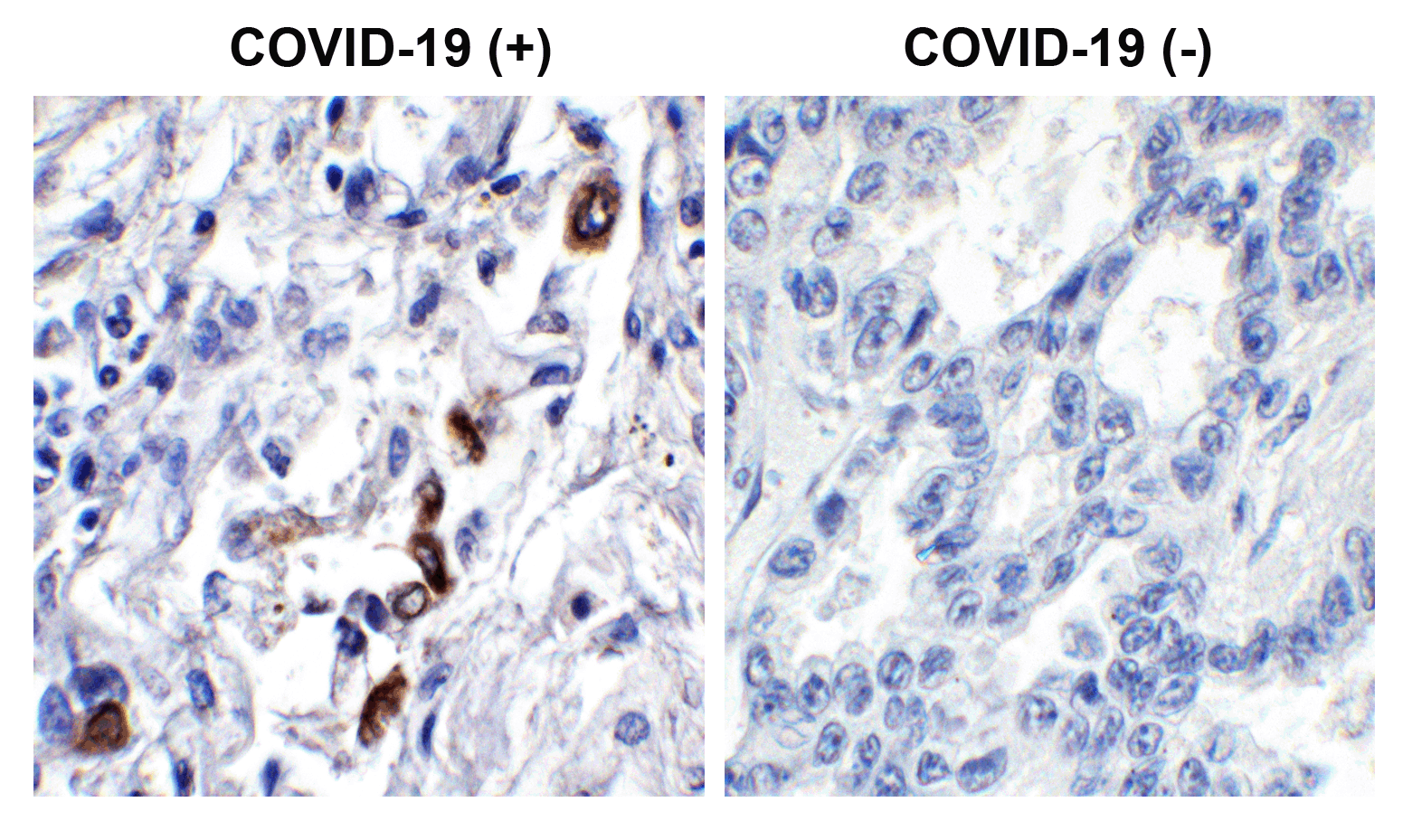
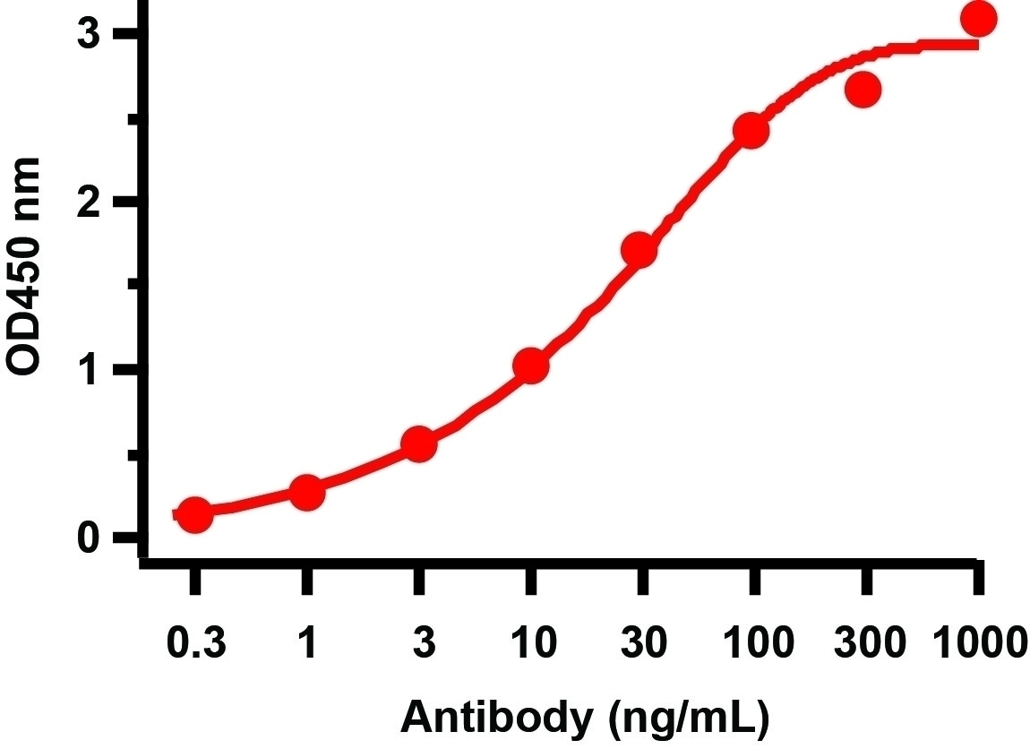
- 产品详情
- 实验流程
- 背景知识
Application 
| IHC, E |
|---|---|
| Primary Accession | P0DTC3 |
| Other Accession | P0DTC3 |
| Host | Rabbit |
| Clonality | Polyclonal |
| Isotype | IgG |
| Clone Names | ORF3a |
| Calculated MW | 31123 Da |
| Concentration (mg/ml) | 1 mg/mL |
| Conjugate | Unconjugated |
| Application Notes | IHC: 0.2 μg/mL Antibody validated: Immunohistochemistry in human samples. Antibody validated: SARS-CoV-2 (COVID-19) ORF3a antibody can detect 2 ng of free peptide at 1 μg/mL in ELISA. All other applications and species not yet tested. |
| Gene ID | 43740569 |
|---|---|
| Other Names | ORF3a protein, Accessory protein 3a, Protein 3a, Protein U274, Protein X1, ORF3a |
| Reconstitution & Storage | SARS-CoV-2 (COVID-19) ORF3a antibody can be stored at 4˚C for three months and -20˚C, stable for up to one year. As with all antibodies care should be taken to avoid repeated freeze thaw cycles. Antibodies should not be exposed to prolonged high temperatures. |
| Precautions | SARS-CoV-2 (COVID-19) ORF3a Antibody is for research use only and not for use in diagnostic or therapeutic procedures. |
For Research Use Only. Not For Use In Diagnostic Procedures.
| Name | 3a |
|---|---|
| Function | Plays a role in viral egress via lysosomal trafficking (PubMed:33157038, PubMed:33422265). Forms homotetrameric ion channels (viroporins) localized at endosomes and lysosomes, that may induce deacidification of lysosomes, allowing safe egress of virions via lysosomal trafficking (PubMed:33157038, PubMed:33422265, PubMed:34158638). Also blocks autolysosome formation by binding and sequestering the host component VPS39 for homotypic fusion and protein sorting (HOPS) on late endosomes (PubMed:33422265). This prevents fusion of autophagosomes with lysosomes, disrupting autophagy and facilitating virus egress (PubMed:33422265). Induces host RETREG1/FAM134B-dependent reticulophagy by interacting with host HMGB1 and enhancing the association between HMGB1 and host BECN1 (PubMed:35239449). This induces endoplasmic reticulum stress and inflammatory responses and facilitates viral infection (PubMed:35239449). |
| Cellular Location | Virion {ECO:0000250|UniProtKB:P59632}. Host cell membrane {ECO:0000250|UniProtKB:P59632, ECO:0000269|PubMed:33060197, ECO:0000269|PubMed:34158638}; Multi-pass membrane protein {ECO:0000250|UniProtKB:P59632, ECO:0000269|PubMed:34158638}. Host endoplasmic reticulum membrane; Multi- pass membrane protein. Secreted {ECO:0000250|UniProtKB:P59632}. Host cytoplasm {ECO:0000250|UniProtKB:P59632, ECO:0000269|PubMed:33060197}. Host endosome Host lysosome. Note=The cell surface expressed protein can undergo endocytosis. The protein is secreted in association with membranous structures. {ECO:0000250|UniProtKB:P59632} |
Provided below are standard protocols that you may find useful for product applications.
BACKGROUND
Coronavirus disease 2019 (COVID-19), formerly known as 2019-nCoV acute respiratory disease, is an infectious disease caused by SARS-CoV-2, a virus closely related to the SARS virus (1). The disease is the cause of the 2019–20 coronavirus outbreak (2). SARS-CoV-2 virus proteins include structural proteins, non-structural proteins and accessory factors. The structure of SARS-CoV-2 consists of the following: a spike protein (S), hemagglutinin-esterease dimer (HE), a membrane glycoprotein (M), an envelope protein (E) a nucleoclapid protein (N) and RNA. SARS-CoV-2 non-structural protein is ORF1ab that consists of 16 proteins (nsp1-nsp16), while accessory factors include ORF3a, ORF3b, ORF6, ORF7a, ORF7b, ORF8, ORF9b, ORF9c and ORF10. ORF3a forms homotetrameric potassium sensitive ion channels (viroporin) and may modulate virus release. It up-regulates expression of fibrinogen subunits FGA, FGB and FGG in host lung epithelial cells. It induces apoptosis in cell culture and downregulates the type 1 interferon receptor by inducing serine phosphorylation within the IFN alpha-receptor subunit 1 (IFNAR1) degradation motif and increasing IFNAR1 ubiquitination (3).
REFERENCES
Gorbalenya. bioRxiv: 2020.;Hui et al. Int J Infect Dis. 2020;91:264-266.;Lu et al. PNAS USA. 2006; 103:12540-12545.

终于等到您。ABCEPTA(百远生物)抗体产品。
点击下方“我要评价 ”按钮提交您的反馈信息,您的反馈和评价是我们最宝贵的财富之一,
我们将在1-3个工作日内处理您的反馈信息。
如有疑问,联系:0512-88856768 tech-china@abcepta.com.


