


 癌症的基本特征包括细胞增殖、血管生成、迁移、凋亡逃避机制和细胞永生等。找到癌症发生过程中这些通路的关键标记物和对应的抗体用于检测至关重要。
 为您推荐一个泛素化位点预测神器——泛素化分析工具,可以为您的蛋白的泛素化位点作出预测和评分。
 细胞自噬受体图形绘图工具为你的蛋白的细胞受体结合位点作出预测和评分,识别结合到自噬通路中的蛋白是非常重要的,便于让我们理解自噬在正常生理、病理过程中的作用,如发育、细胞分化、神经退化性疾病、压力条件下、感染和癌症。
Anti-Rat IgG (H&L) (Biotin Conjugated) Secondary Antibody
Goat Polyclonal, Biotin
- 产品详情
- 实验流程
- 背景知识
| Description | Anti-RAT IgG (H&L) (GOAT) Antibody Biotin Conjugated |
|---|---|
| Host | Goat |
| Conjugate | Biotin |
| Target Species | Rat |
| Clonality | Polyclonal |
Application
| WB |
| Physical State | Lyophilized |
| Host Isotype | IgG |
| Target Isotype | IgG (H&L) |
| Buffer | 0.02 M Potassium Phosphate, 0.15 M Sodium Chloride, pH 7.2 |
| Immunogen | Rat IgG whole molecule |
| Reconstitution Volume | 1.0 mL |
| Reconstitution Buffer | Restore with deionized water (or equivalent) |
| Stabilizer | 10 mg/mL Bovine Serum Albumin (BSA) - Immunoglobulin and Protease free |
| Preservative | 0.01% (w/v) Sodium Azide |
| Shipping Condition | Ambient |
|---|---|
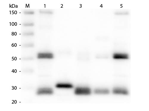
| Application Note | Anti-RAT IgG Biotin Conjugated antibody is suitable for immunoassays where specificity to the Rat immunoglobulin heavy and or light chain regions is desired. Anti-Rat antibody has been assayed against 1.0 µg of Rat IgG in a standard capture ELISA using Peroxidase Conjugated Streptavidin and ABTS (2,2’-azino-bis-[3-ethylbenthiazoline-6-sulfonic acid]) as a substrate for 30 minutes at room temperature. A working dilution of 1:100,000 to 1:500,000 is suggested for this product. Optimal concentrations in immunoassays should be determined by the researcher. |
| Purity | Anti-RAT IgG Biotin Conjugated antibody was prepared from monospecific antiserum by immunoaffinity chromatography using Rat IgG coupled to agarose. Assay by immunoelectrophoresis resulted in a single precipitin arc against anti-biotin, anti-Goat Serum, Rat IgG and Rat Serum. |
| Storage Condition | Store vial at 4° C prior to restoration. For extended storage aliquot contents and freeze at -20° C or below. Avoid cycles of freezing and thawing. Centrifuge product if not completely clear after standing at room temperature. This product is stable for several weeks at 4° C as an undiluted liquid. Dilute only prior to immediate use. |
| Precautions Note | This product is for research use only and is not intended for therapeutic or diagnostic applications. |
For Research Use Only. Not For Use In Diagnostic Procedures.
Provided below are standard protocols that you may find useful for product applications.
BACKGROUND
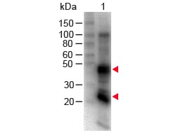
RAT IgG Biotin Conjugated antibody detects rat immunoglobulin G. Immunoglobuoin G is a molecule of about 150 kDa composed of four peptide chains. Each IgG contains two identical ? heavy chains of about 51 kDa and two identical light chains of about 26 kDa, thus a tetrameric quaternary structure. The two heavy chains are linked to each other and to a light chain each by disulfide bonds. The resulting tetramer has two identical halves, which together form the Y-like shape. Each end of the fork contains an identical antigen binding site. The Fc regions of IgGs bear a highly conserved N-glycosylation site.
终于等到您。ABCEPTA(百远生物)抗体产品。
点击下方“我要评价 ”按钮提交您的反馈信息,您的反馈和评价是我们最宝贵的财富之一,
我们将在1-3个工作日内处理您的反馈信息。
如有疑问,联系:0512-88856768 tech-china@abcepta.com.


