


 癌症的基本特征包括细胞增殖、血管生成、迁移、凋亡逃避机制和细胞永生等。找到癌症发生过程中这些通路的关键标记物和对应的抗体用于检测至关重要。
 为您推荐一个泛素化位点预测神器——泛素化分析工具,可以为您的蛋白的泛素化位点作出预测和评分。
 细胞自噬受体图形绘图工具为你的蛋白的细胞受体结合位点作出预测和评分,识别结合到自噬通路中的蛋白是非常重要的,便于让我们理解自噬在正常生理、病理过程中的作用,如发育、细胞分化、神经退化性疾病、压力条件下、感染和癌症。
Anti-Rabbit IgG (H&L) (ATTO 425 Conjugated) Pre-Adsorbed Secondary Antibody
Goat Polyclonal, ATTO 425
- 产品详情
- 实验流程
- 背景知识
| Description | Anti-RABBIT IgG (H&L) (GOAT) Antibody ATTO 425 Conjugated (Min X Bv Ch Gt GP Ham Hs Hu Ms Rt & Sh Serum Proteins) |
|---|---|
| Host | Goat |
| Conjugate | ATTO 425 |
| FP Value | 2.3 moles ATTO 425 per mole of IgG |
| Target Species | Rabbit |
| Clonality | Polyclonal |
Application
| IF, WB |
| Physical State | Lyophilized |
| Host Isotype | IgG |
| Target Isotype | IgG (H&L) |
| Buffer | 0.02 M Potassium Phosphate, 0.15 M Sodium Chloride, pH 7.2 |
| Immunogen | Rabbit IgG whole molecule |
| Reconstitution Volume | 500 µL |
| Reconstitution Buffer | Restore with deionized water (or equivalent) |
| Stabilizer | 10 mg/mL Bovine Serum Albumin (BSA) - Immunoglobulin and Protease free |
| Preservative | 0.01% (w/v) Sodium Azide |
| Shipping Condition | Ambient |
|---|---|
| Application Note | The emission spectra for this ATTO conjugate matches the principle output wavelengths of most common fluorescence instrumentation. |
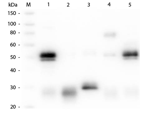
| Purity | Rabbit IgG (H&L) Antibody ATTO 425 was prepared from monospecific antiserum by immunoaffinity chromatography using Rabbit IgG coupled to agarose beads followed by solid phase adsorption(s) to remove any unwanted reactivities. Assay by immunoelectrophoresis resulted in a single precipitin arc against anti-Goat Serum, Rabbit IgG and Rabbit Serum. No reaction was observed against Bovine, Chicken, Goat, Guinea Pig, Hamster, Horse, Human, Mouse, Rat and Sheep Serum Proteins. This antibody will react with heavy chains of rabbit IgG and with light chains of most rabbit immunoglobulins. |
| Storage Condition | Store vial at 4° C prior to restoration. For extended storage aliquot contents and freeze at -20° C or below. Avoid cycles of freezing and thawing. Centrifuge product if not completely clear after standing at room temperature. This product is stable for several weeks at 4° C as an undiluted liquid. Dilute only prior to immediate use. |
| Precautions Note | This product is for research use only and is not intended for therapeutic or diagnostic applications. |
Research Areas
For Research Use Only. Not For Use In Diagnostic Procedures.
Application Protocols
Provided below are standard protocols that you may find useful for product applications.
BACKGROUND
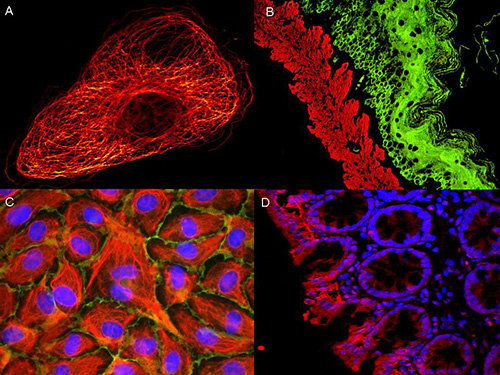
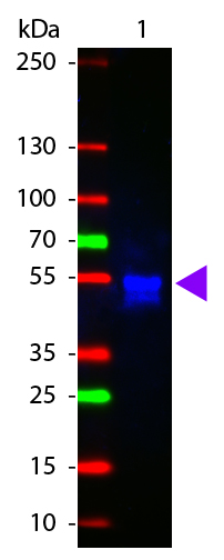
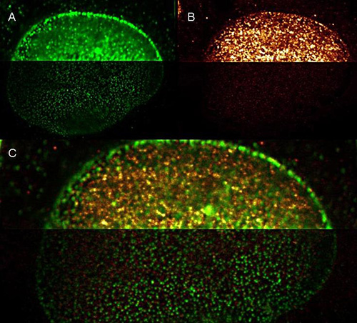
Anti-Rabbit IgG (H&L) conjugated to ATTO 425 is designed for STED microscopy, FRET, immunofluorescence microscopy, fluorescence based plate assays (FLISA) and fluorescent western blotting. This product is also suitable for multiplex analysis, including multicolor imaging, utilizing various commercial platforms.
终于等到您。ABCEPTA(百远生物)抗体产品。
点击下方“我要评价 ”按钮提交您的反馈信息,您的反馈和评价是我们最宝贵的财富之一,
我们将在1-3个工作日内处理您的反馈信息。
如有疑问,联系:0512-88856768 tech-china@abcepta.com.


