


 癌症的基本特征包括细胞增殖、血管生成、迁移、凋亡逃避机制和细胞永生等。找到癌症发生过程中这些通路的关键标记物和对应的抗体用于检测至关重要。
 为您推荐一个泛素化位点预测神器——泛素化分析工具,可以为您的蛋白的泛素化位点作出预测和评分。
 细胞自噬受体图形绘图工具为你的蛋白的细胞受体结合位点作出预测和评分,识别结合到自噬通路中的蛋白是非常重要的,便于让我们理解自噬在正常生理、病理过程中的作用,如发育、细胞分化、神经退化性疾病、压力条件下、感染和癌症。
HYOU1 Antibody (monoclonal) (M01)
Mouse monoclonal antibody raised against a partial recombinant HYOU1.
- 产品详情
- 实验流程
- 背景知识
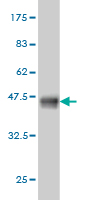
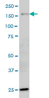
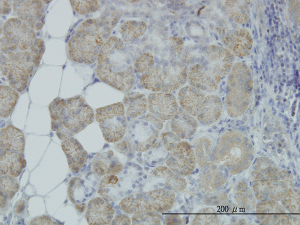
Application
| WB, IHC, E |
|---|---|
| Primary Accession | Q9Y4L1 |
| Other Accession | NM_006389 |
| Reactivity | Human |
| Host | mouse |
| Clonality | monoclonal |
| Isotype | IgG1 Kappa |
| Clone Names | 6F7 |
| Calculated MW | 111335 Da |
| Gene ID | 10525 |
|---|---|
| Other Names | Hypoxia up-regulated protein 1, 150 kDa oxygen-regulated protein, ORP-150, 170 kDa glucose-regulated protein, GRP-170, HYOU1, GRP170, ORP150 |
| Target/Specificity | HYOU1 (NP_006380, 901 a.a. ~ 999 a.a) partial recombinant protein with GST tag. MW of the GST tag alone is 26 KDa. |
| Dilution | WB~~1:500~1000 IHC~~1:100~500 E~~N/A |
| Format | Clear, colorless solution in phosphate buffered saline, pH 7.2 . |
| Storage | Store at -20°C or lower. Aliquot to avoid repeated freezing and thawing. |
| Precautions | HYOU1 Antibody (monoclonal) (M01) is for research use only and not for use in diagnostic or therapeutic procedures. |
For Research Use Only. Not For Use In Diagnostic Procedures.
Provided below are standard protocols that you may find useful for product applications.
BACKGROUND
The protein encoded by this gene belongs to the heat shock protein 70 family. This gene uses alternative transcription start sites. A cis-acting segment found in the 5' UTR is involved in stress-dependent induction, resulting in the accumulation of this protein in the endoplasmic reticulum (ER) under hypoxic conditions. The protein encoded by this gene is thought to play an important role in protein folding and secretion in the ER. Since suppression of the protein is associated with accelerated apoptosis, it is also suggested to have an important cytoprotective role in hypoxia-induced cellular perturbation. This protein has been shown to be up-regulated in tumors, especially in breast tumors, and thus it is associated with tumor invasiveness. This gene also has an alternative translation initiation site, resulting in a protein that lacks the N-terminal signal peptide. This signal peptide-lacking protein, which is only 3 amino acids shorter than the mature protein in the ER, is thought to have a housekeeping function in the cytosol. In rat, this protein localizes to both the ER by a carboxy-terminal peptide sequence and to mitochondria by an amino-terminal targeting signal.
REFERENCES
1.Limited expression of reticulocalbin-1 in lymphatic endothelial cells in lung tumor but not in normal lung.Yoshida Y, Yamashita T, Nagano K, Imai S, Nabeshi H, Yoshikawa T, Yoshioka Y, Abe Y, Kamada H, Tsutsumi Y, Tsunoda SI.Biochem Biophys Res Commun. 2011 Jan 25. [Epub ahead of print]2.Proteinuria and Hyperglycemia Induce Endoplasmic Reticulum Stress.Lindenmeyer MT, Rastaldi MP, Ikehata M, Neusser MA, Kretzler M, Cohen CD, Schlondorff D.J Am Soc Nephrol. 2008 Nov;19(11):2225-36. Epub 2008 Sep 5.3.Mechanism of cancer cell adaptation to metabolic stress: proteomics identification of a novel thyroid hormone mediated gastric carcinogenic signaling pathway.Liu R, Li Z, Bai S, Zhang H, Tang M, Lei Y, Chen L, Liang S, Zhao YL, Wei Y, Huang C.Mol Cell Proteomics. 2009 Jan;8(1):70-85. Epub 2008 Aug 22.
终于等到您。ABCEPTA(百远生物)抗体产品。
点击下方“我要评价 ”按钮提交您的反馈信息,您的反馈和评价是我们最宝贵的财富之一,
我们将在1-3个工作日内处理您的反馈信息。
如有疑问,联系:0512-88856768 tech-china@abcepta.com.


