


 癌症的基本特征包括细胞增殖、血管生成、迁移、凋亡逃避机制和细胞永生等。找到癌症发生过程中这些通路的关键标记物和对应的抗体用于检测至关重要。
 为您推荐一个泛素化位点预测神器——泛素化分析工具,可以为您的蛋白的泛素化位点作出预测和评分。
 细胞自噬受体图形绘图工具为你的蛋白的细胞受体结合位点作出预测和评分,识别结合到自噬通路中的蛋白是非常重要的,便于让我们理解自噬在正常生理、病理过程中的作用,如发育、细胞分化、神经退化性疾病、压力条件下、感染和癌症。
KRT4 Antibody (monoclonal) (M01)
Mouse monoclonal antibody raised against a partial recombinant KRT4.
- 产品详情
- 实验流程
- 背景知识
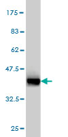
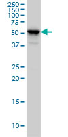
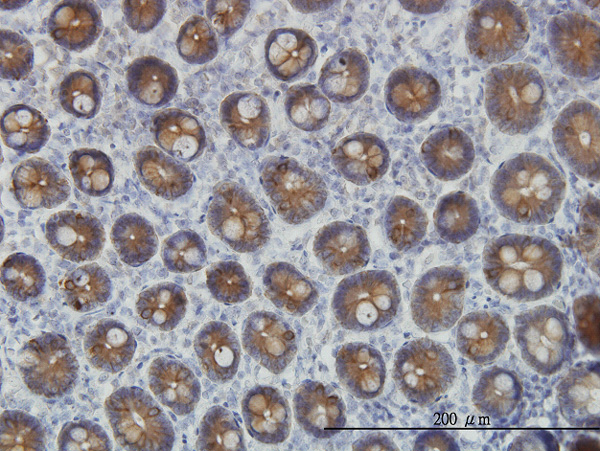
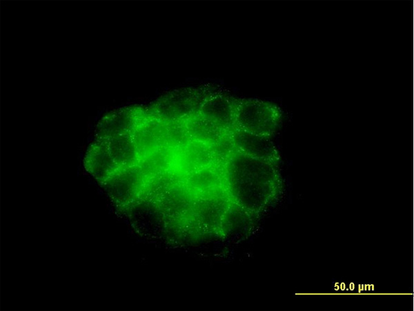
Application
| WB, IHC, IF, E |
|---|---|
| Primary Accession | P19013 |
| Other Accession | NM_002272 |
| Reactivity | Human |
| Host | mouse |
| Clonality | monoclonal |
| Isotype | IgG1 Kappa |
| Clone Names | 5H5 |
| Calculated MW | 56144 Da |
| Gene ID | 3851 |
|---|---|
| Other Names | Keratin, type II cytoskeletal 4, Cytokeratin-4, CK-4, Keratin-4, K4, Type-II keratin Kb4, KRT4, CYK4 |
| Target/Specificity | KRT4 (NP_002263, 194 a.a. ~ 300 a.a) partial recombinant protein with GST tag. MW of the GST tag alone is 26 KDa. |
| Dilution | WB~~1:500~1000 IHC~~1:100~500 IF~~1:50~200 E~~N/A |
| Format | Clear, colorless solution in phosphate buffered saline, pH 7.2 . |
| Storage | Store at -20°C or lower. Aliquot to avoid repeated freezing and thawing. |
| Precautions | KRT4 Antibody (monoclonal) (M01) is for research use only and not for use in diagnostic or therapeutic procedures. |
For Research Use Only. Not For Use In Diagnostic Procedures.
Provided below are standard protocols that you may find useful for product applications.
BACKGROUND
The protein encoded by this gene is a member of the keratin gene family. The type II cytokeratins consist of basic or neutral proteins which are arranged in pairs of heterotypic keratin chains coexpressed during differentiation of simple and stratified epithelial tissues. This type II cytokeratin is specifically expressed in differentiated layers of the mucosal and esophageal epithelia with family member KRT13. Mutations in these genes have been associated with White Sponge Nevus, characterized by oral, esophageal, and anal leukoplakia. The type II cytokeratins are clustered in a region of chromosome 12q12-q13.
REFERENCES
Defining the human deubiquitinating enzyme interaction landscape. Sowa ME, et al. Cell, 2009 Jul 23. PMID 19615732.Comparative analysis of the expression of cytokeratins (1, 10, 14, 16, 4), involucrin, filaggrin and e-cadherin in plane warts and epidermodysplasia verruciformis plane wart-type lesions. Barcelos AC, et al. J Cutan Pathol, 2009 Jun. PMID 19515043.Plk1 self-organization and priming phosphorylation of HsCYK-4 at the spindle midzone regulate the onset of division in human cells. Burkard ME, et al. PLoS Biol, 2009 May 5. PMID 19468302.Polo-like kinase 1 directs assembly of the HsCyk-4 RhoGAP/Ect2 RhoGEF complex to initiate cleavage furrow formation. Wolfe BA, et al. PLoS Biol, 2009 May 5. PMID 19468300.Two new mutations in the keratin 4 gene causing oral white sponge nevus in Chinese family. Zhang JM, et al. Oral Dis, 2009 Jan. PMID 18992023.
终于等到您。ABCEPTA(百远生物)抗体产品。
点击下方“我要评价 ”按钮提交您的反馈信息,您的反馈和评价是我们最宝贵的财富之一,
我们将在1-3个工作日内处理您的反馈信息。
如有疑问,联系:0512-88856768 tech-china@abcepta.com.


