


 癌症的基本特征包括细胞增殖、血管生成、迁移、凋亡逃避机制和细胞永生等。找到癌症发生过程中这些通路的关键标记物和对应的抗体用于检测至关重要。
 为您推荐一个泛素化位点预测神器——泛素化分析工具,可以为您的蛋白的泛素化位点作出预测和评分。
 细胞自噬受体图形绘图工具为你的蛋白的细胞受体结合位点作出预测和评分,识别结合到自噬通路中的蛋白是非常重要的,便于让我们理解自噬在正常生理、病理过程中的作用,如发育、细胞分化、神经退化性疾病、压力条件下、感染和癌症。
UBE2K Antibody(N-term)
Affinity Purified Rabbit Polyclonal Antibody (Pab)

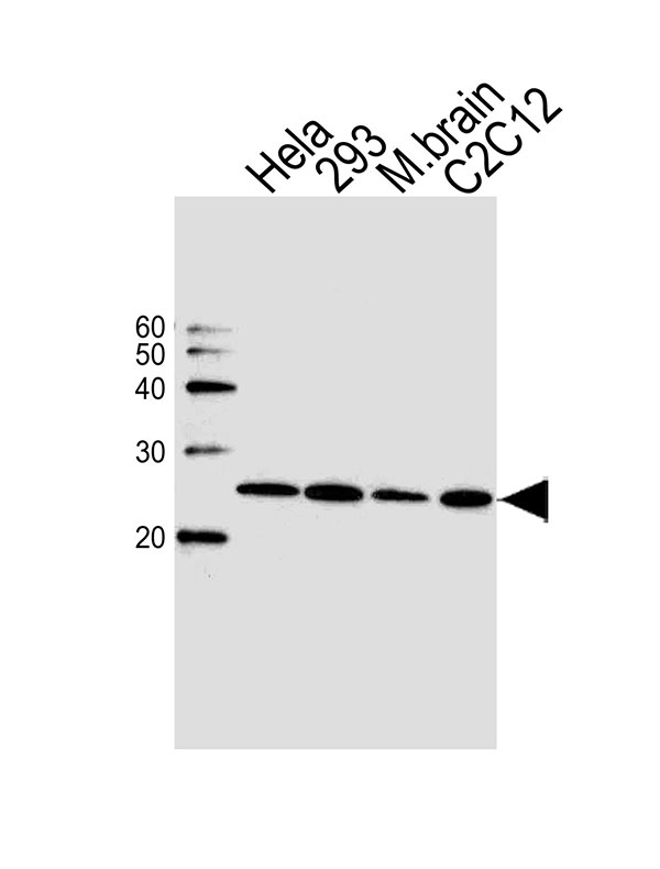
- 产品详情
- 实验流程
- 背景知识
Application 
| WB |
|---|---|
| Primary Accession | P61086 |
| Other Accession | P61087, P61085, NP_005330.1 |
| Reactivity | Mouse, Human |
| Predicted | Bovine |
| Host | Rabbit |
| Clonality | Polyclonal |
| Calculated MW | 22407 Da |
| Isotype | Rabbit IgG |
| Antigen Source | HUMAN |
| Gene ID | 3093 |
|---|---|
| Antigen Region | 2-30 aa |
| Other Names | UBE2K; HIP2; LIG; Ubiquitin-conjugating enzyme E2 K; Huntingtin-interacting protein 2; Ubiquitin carrier protein; Ubiquitin-conjugating enzyme E2-25 kDa; Ubiquitin-protein ligase |
| Dilution | WB~~1:500 |
| Target/Specificity | This HIP2 (UBE2K) antibody is generated from rabbits immunized with a KLH conjugated synthetic peptide between 2-30 amino acids from the N-terminal region of human UBE2K. |
| Format | Purified polyclonal antibody supplied in PBS with 0.09% (W/V) sodium azide. This antibody is purified through a protein A column, followed by peptide affinity purification. |
| Storage | Maintain refrigerated at 2-8°C for up to 2 weeks. For long term storage store at -20°C in small aliquots to prevent freeze-thaw cycles. |
| Precautions | UBE2K Antibody(N-term) is for research use only and not for use in diagnostic or therapeutic procedures. |
For Research Use Only. Not For Use In Diagnostic Procedures.
| Name | UBE2K |
|---|---|
| Synonyms | HIP2, LIG |
| Function | Accepts ubiquitin from the E1 complex and catalyzes its covalent attachment to other proteins. In vitro, in the presence or in the absence of BRCA1-BARD1 E3 ubiquitin-protein ligase complex, catalyzes the synthesis of 'Lys-48'-linked polyubiquitin chains. Does not transfer ubiquitin directly to but elongates monoubiquitinated substrate protein. Mediates the selective degradation of short-lived and abnormal proteins, such as the endoplasmic reticulum-associated degradation (ERAD) of misfolded lumenal proteins. Ubiquitinates huntingtin. May mediate foam cell formation by the suppression of apoptosis of lipid-bearing macrophages through ubiquitination and subsequence degradation of p53/TP53. Proposed to be involved in ubiquitination and proteolytic processing of NF-kappa-B; in vitro supports ubiquitination of NFKB1. In case of infection by cytomegaloviruses may be involved in the US11-dependent degradation of MHC class I heavy chains following their export from the ER to the cytosol. In case of viral infections may be involved in the HPV E7 protein-dependent degradation of RB1. |
| Cellular Location | Cytoplasm {ECO:0000250|UniProtKB:P61085}. |
| Tissue Location | Expressed in all tissues tested, including spleen, thymus, prostate, testis, ovary, small intestine, colon, peripheral blood leukocytes, T-lymphocytes, monocytes, granulocytes and bone marrow mononuclear cells. Highly expressed in brain, with highest levels found in cortex and striatum and at lower levels in cerebellum and brainstem. |
Provided below are standard protocols that you may find useful for product applications.
BACKGROUND
The protein encoded by this gene belongs to the ubiquitin-conjugating enzyme family. This protein interacts with RING finger proteins, and it can ubiquitinate huntingtin, the gene product for Huntington's disease. Known functions for this protein include a role in aggregate formation of expanded polyglutamine proteins and the suppression of apoptosis in polyglutamine diseases, a role in the dislocation of newly synthesized MHC class I heavy chains from the endoplasmic reticulum, and involvement in foam cell formation. Multiple transcript variants encoding different isoforms have been identified for this gene. [provided by RefSeq].
REFERENCES
Bae, Y., et al. Biochem. Biophys. Res. Commun. 397(4):718-723(2010)
Christensen, D.E., et al. Nat. Struct. Mol. Biol. 14(10):941-948(2007)
de Pril, R., et al. Mol. Cell. Neurosci. 34(1):10-19(2007)
Flierman, D., et al. Proc. Natl. Acad. Sci. U.S.A. 103(31):11589-11594(2006)
Yamada, M., et al. J. Biol. Chem. 281(30):20749-20760(2006)

终于等到您。ABCEPTA(百远生物)抗体产品。
点击下方“我要评价 ”按钮提交您的反馈信息,您的反馈和评价是我们最宝贵的财富之一,
我们将在1-3个工作日内处理您的反馈信息。
如有疑问,联系:0512-88856768 tech-china@abcepta.com.


