


 癌症的基本特征包括细胞增殖、血管生成、迁移、凋亡逃避机制和细胞永生等。找到癌症发生过程中这些通路的关键标记物和对应的抗体用于检测至关重要。
 为您推荐一个泛素化位点预测神器——泛素化分析工具,可以为您的蛋白的泛素化位点作出预测和评分。
 细胞自噬受体图形绘图工具为你的蛋白的细胞受体结合位点作出预测和评分,识别结合到自噬通路中的蛋白是非常重要的,便于让我们理解自噬在正常生理、病理过程中的作用,如发育、细胞分化、神经退化性疾病、压力条件下、感染和癌症。
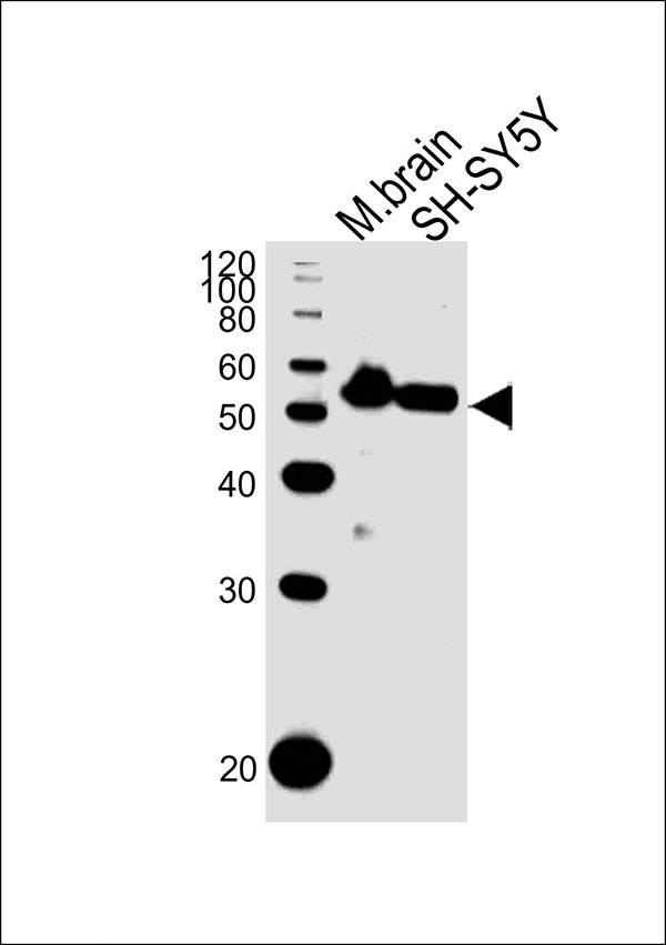
DAPK2 Antibody (N-term)
Purified Rabbit Polyclonal Antibody (Pab)
- 产品详情
- 实验流程
- 背景知识
Application
| WB |
|---|---|
| Primary Accession | Q9UIK4 |
| Reactivity | Mouse, Human |
| Host | Rabbit |
| Clonality | Polyclonal |
| Calculated MW | 42898 Da |
| Isotype | Rabbit IgG |
| Antigen Source | HUMAN |
| Gene ID | 23604 |
|---|---|
| Antigen Region | 1-30 aa |
| Other Names | Death-associated protein kinase 2, DAP kinase 2, DAP-kinase-related protein 1, DRP-1, DAPK2 |
| Dilution | WB~~1:1000 |
| Target/Specificity | This DAPK2 antibody is generated from rabbits immunized with a KLH conjugated synthetic peptide between 1-30 amino acids from the N-terminal region of human DAPK2. |
| Format | Purified polyclonal antibody supplied in PBS with 0.09% (W/V) sodium azide. This antibody is purified through a protein A column, followed by peptide affinity purification. |
| Storage | Maintain refrigerated at 2-8°C for up to 2 weeks. For long term storage store at -20°C in small aliquots to prevent freeze-thaw cycles. |
| Precautions | DAPK2 Antibody (N-term) is for research use only and not for use in diagnostic or therapeutic procedures. |
| Name | DAPK2 |
|---|---|
| Function | Calcium/calmodulin-dependent serine/threonine kinase involved in multiple cellular signaling pathways that trigger cell survival, apoptosis, and autophagy. Regulates both type I apoptotic and type II autophagic cell death signals, depending on the cellular setting. The former is caspase-dependent, while the latter is caspase-independent and is characterized by the accumulation of autophagic vesicles. Acts as a mediator of anoikis and a suppressor of beta-catenin-dependent anchorage-independent growth of malignant epithelial cells. May play a role in granulocytic maturation (PubMed:17347302). Regulates granulocytic motility by controlling cell spreading and polarization (PubMed:24163421). |
| Cellular Location | Cytoplasm. Cytoplasmic vesicle, autophagosome lumen |
| Tissue Location | Expressed in neutrophils and eosinophils (PubMed:24163421). Isoform 2 is expressed in embryonic stem cells (at protein level). Isoform 1 is ubiquitously expressed in all tissue types examined with high levels in heart, lung and skeletal muscle |
For Research Use Only. Not For Use In Diagnostic Procedures.
Provided below are standard protocols that you may find useful for product applications.
BACKGROUND
DAPK2 belongs to the serine/threonine protein kinase family. This protein contains a N-terminal protein kinase domain followed by a conserved calmodulin-binding domain with significant similarity to that of death-associated protein kinase 1 (DAPK1), a positive regulator of programmed cell death. Overexpression of this gene was shown to induce cell apoptosis. It uses multiple polyadenylation sites.
REFERENCES
Satoh, A., et al., Br. J. Cancer 86(11):1817-1823 (2002).
Chan, M.W., et al., Clin. Cancer Res. 8(2):464-470 (2002).
Wong, T.S., et al., Clin. Cancer Res. 8(2):433-437 (2002).
Shani, G., et al., EMBO J. 20(5):1099-1113 (2001).
Inbal, B., et al., Mol. Cell. Biol. 20(3):1044-1054 (2000).
终于等到您。ABCEPTA(百远生物)抗体产品。
点击下方“我要评价 ”按钮提交您的反馈信息,您的反馈和评价是我们最宝贵的财富之一,
我们将在1-3个工作日内处理您的反馈信息。
如有疑问,联系:0512-88856768 tech-china@abcepta.com.


