


 癌症的基本特征包括细胞增殖、血管生成、迁移、凋亡逃避机制和细胞永生等。找到癌症发生过程中这些通路的关键标记物和对应的抗体用于检测至关重要。
 为您推荐一个泛素化位点预测神器——泛素化分析工具,可以为您的蛋白的泛素化位点作出预测和评分。
 细胞自噬受体图形绘图工具为你的蛋白的细胞受体结合位点作出预测和评分,识别结合到自噬通路中的蛋白是非常重要的,便于让我们理解自噬在正常生理、病理过程中的作用,如发育、细胞分化、神经退化性疾病、压力条件下、感染和癌症。
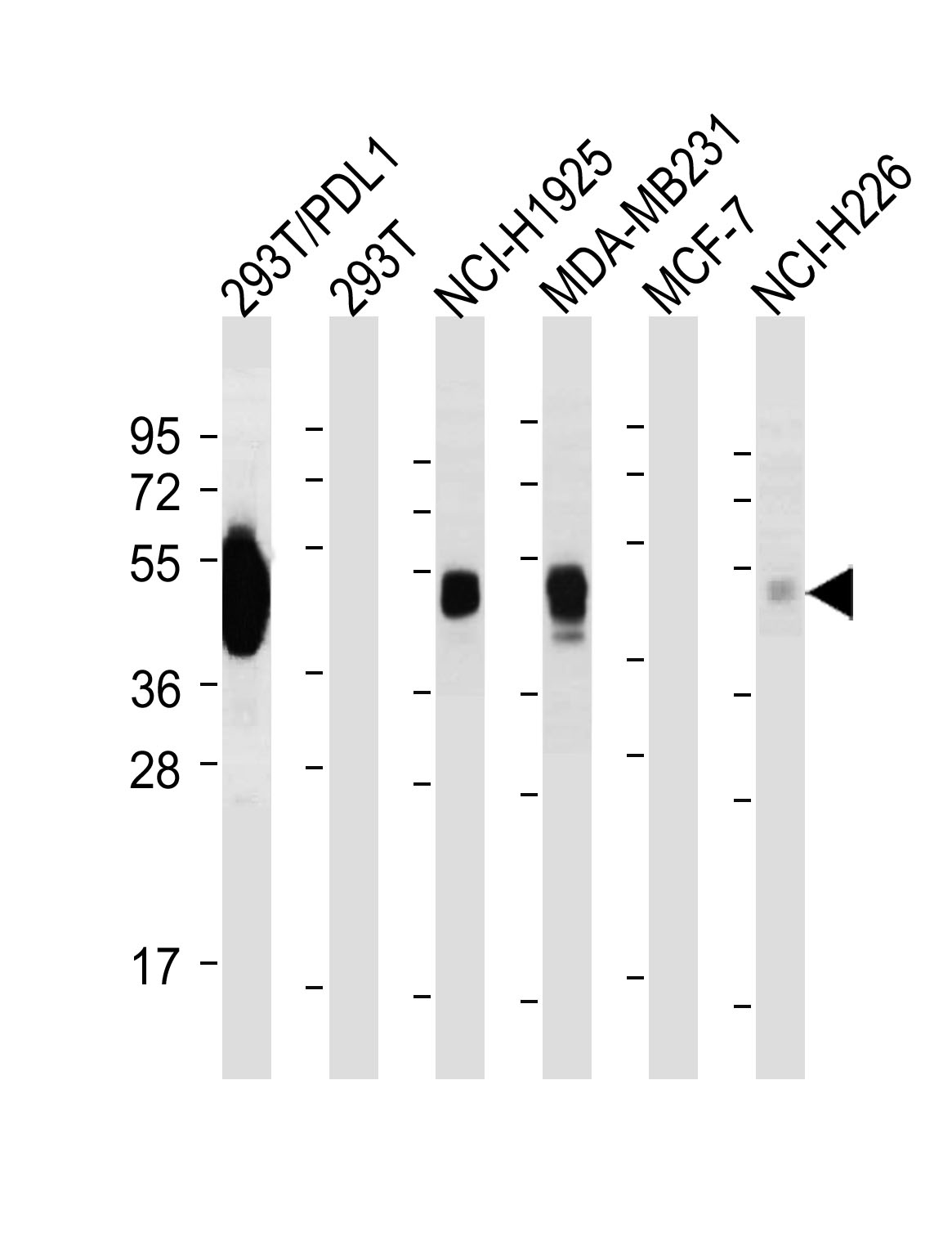
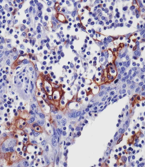
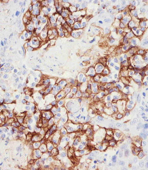
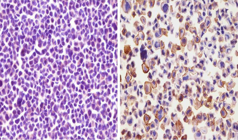
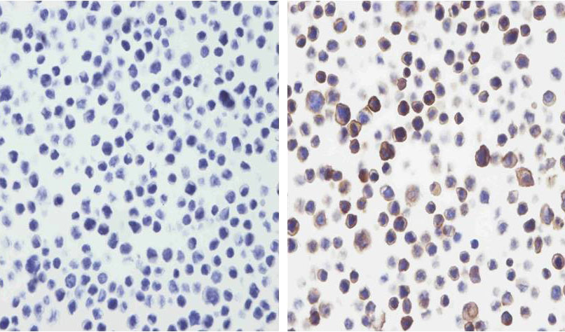
PD L1 Monoclonal Antibody (PDL1)
Purified Mouse Monoclonal Antibody (Mab)
- 产品详情
- 文献引用 : 2
- 实验流程
- 背景知识
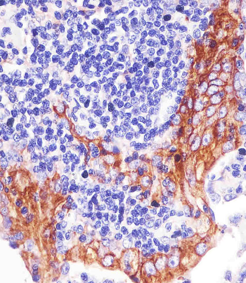
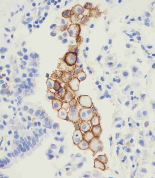
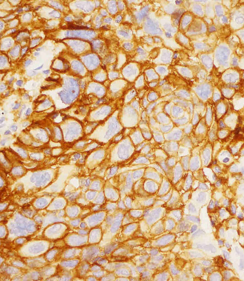
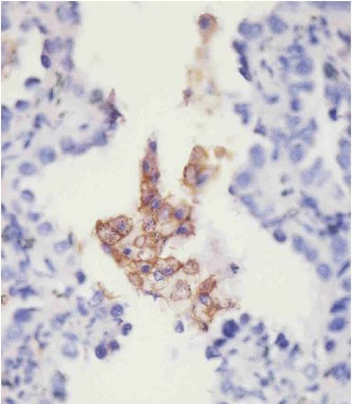
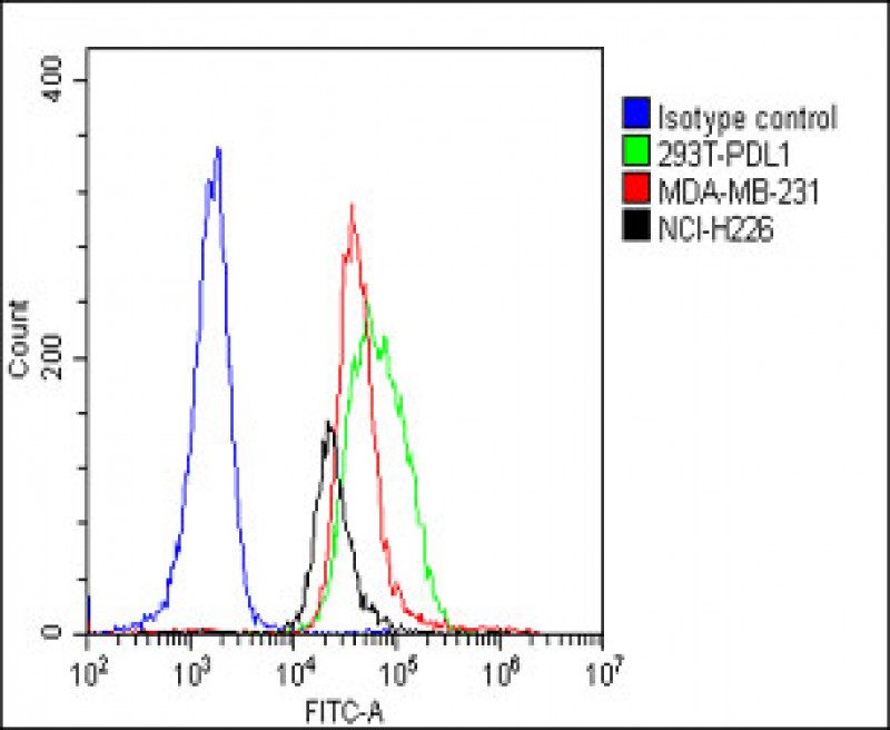
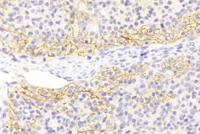
Application
| WB, IHC-P, FC, IHC-P-Leica |
|---|---|
| Primary Accession | Q9NZQ7 |
| Reactivity | Human |
| Predicted | Human |
| Host | Mouse |
| Clonality | monoclonal |
| Calculated MW | 33275 Da |
| Isotype | IgG1 |
| Antigen Source | HUMAN |
| Gene ID | 29126 |
|---|---|
| Other Names | Programmed cell death 1 ligand 1, PD-L1, PDCD1 ligand 1, Programmed death ligand 1, B7 homolog 1, B7-H1, CD274, CD274, B7H1, PDCD1L1, PDCD1LG1, PDL1, PDL-1 |
| Dilution | WB~~1:500-1:1000 IHC-P~~1:500 FC~~1:25 IHC-P-Leica~~1:100-1:600 |
| Target/Specificity | This PD L1 antibody is generated from a mouse immunized with a KLH conjugated synthetic peptide between 256-290 amino acids from the human region of human PD L1. |
| Format | Purified monoclonal antibody supplied in PBS with 0.09% (W/V) sodium azide. This antibody is purified through a protein G column, followed by dialysis against PBS. |
| Storage | Maintain refrigerated at 2-8°C for up to 2 weeks. For long term storage store at -20°C in small aliquots to prevent freeze-thaw cycles. |
| Precautions | PD L1 Monoclonal Antibody (PDL1) is for research use only and not for use in diagnostic or therapeutic procedures. |
| Name | CD274 (HGNC:17635) |
|---|---|
| Function | Plays a critical role in induction and maintenance of immune tolerance to self (PubMed:11015443, PubMed:28813410, PubMed:28813417, PubMed:31399419). As a ligand for the inhibitory receptor PDCD1/PD-1, modulates the activation threshold of T-cells and limits T-cell effector response (PubMed:11015443, PubMed:28813410, PubMed:28813417, PubMed:36727298). Through a yet unknown activating receptor, may costimulate T-cell subsets that predominantly produce interleukin-10 (IL10) (PubMed:10581077). Can also act as a transcription coactivator: in response to hypoxia, translocates into the nucleus via its interaction with phosphorylated STAT3 and promotes transcription of GSDMC, leading to pyroptosis (PubMed:32929201). |
| Cellular Location | Cell membrane; Single-pass type I membrane protein. Early endosome membrane; Single-pass type I membrane protein. Recycling endosome membrane; Single-pass type I membrane protein. Nucleus. Note=Associates with CMTM6 at recycling endosomes, where it is protected from being targeted for lysosomal degradation (PubMed:28813417). Translocates to the nucleus in response to hypoxia via its interaction with phosphorylated STAT3 (PubMed:32929201). [Isoform 2]: Endomembrane system; Single-pass type I membrane protein |
| Tissue Location | Highly expressed in the heart, skeletal muscle, placenta and lung. Weakly expressed in the thymus, spleen, kidney and liver. Expressed on activated T- and B-cells, dendritic cells, keratinocytes and monocytes. |
For Research Use Only. Not For Use In Diagnostic Procedures.

Provided below are standard protocols that you may find useful for product applications.
BACKGROUND
Involved in the costimulatory signal, essential for T- cell proliferation and production of IL10 and IFNG, in an IL2- dependent and a PDCD1-independent manner. Interaction with PDCD1 inhibits T-cell proliferation and cytokine production.
REFERENCES
Dong H.,et al.Nat. Med. 5:1365-1369(1999).
Freeman G.J.,et al.J. Exp. Med. 192:1027-1034(2000).
He X.-H.,et al.Acta Pharmacol. Sin. 26:462-468(2005).
Chi X.-Y.,et al.Submitted (NOV-2005) to the EMBL/GenBank/DDBJ databases.
Ota T.,et al.Nat. Genet. 36:40-45(2004).
终于等到您。ABCEPTA(百远生物)抗体产品。
点击下方“我要评价 ”按钮提交您的反馈信息,您的反馈和评价是我们最宝贵的财富之一,
我们将在1-3个工作日内处理您的反馈信息。
如有疑问,联系:0512-88856768 tech-china@abcepta.com.


