


 癌症的基本特征包括细胞增殖、血管生成、迁移、凋亡逃避机制和细胞永生等。找到癌症发生过程中这些通路的关键标记物和对应的抗体用于检测至关重要。
 为您推荐一个泛素化位点预测神器——泛素化分析工具,可以为您的蛋白的泛素化位点作出预测和评分。
 细胞自噬受体图形绘图工具为你的蛋白的细胞受体结合位点作出预测和评分,识别结合到自噬通路中的蛋白是非常重要的,便于让我们理解自噬在正常生理、病理过程中的作用,如发育、细胞分化、神经退化性疾病、压力条件下、感染和癌症。
Anti-CXCR4 Antibody
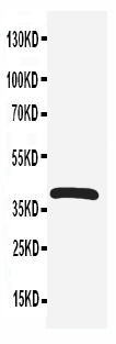
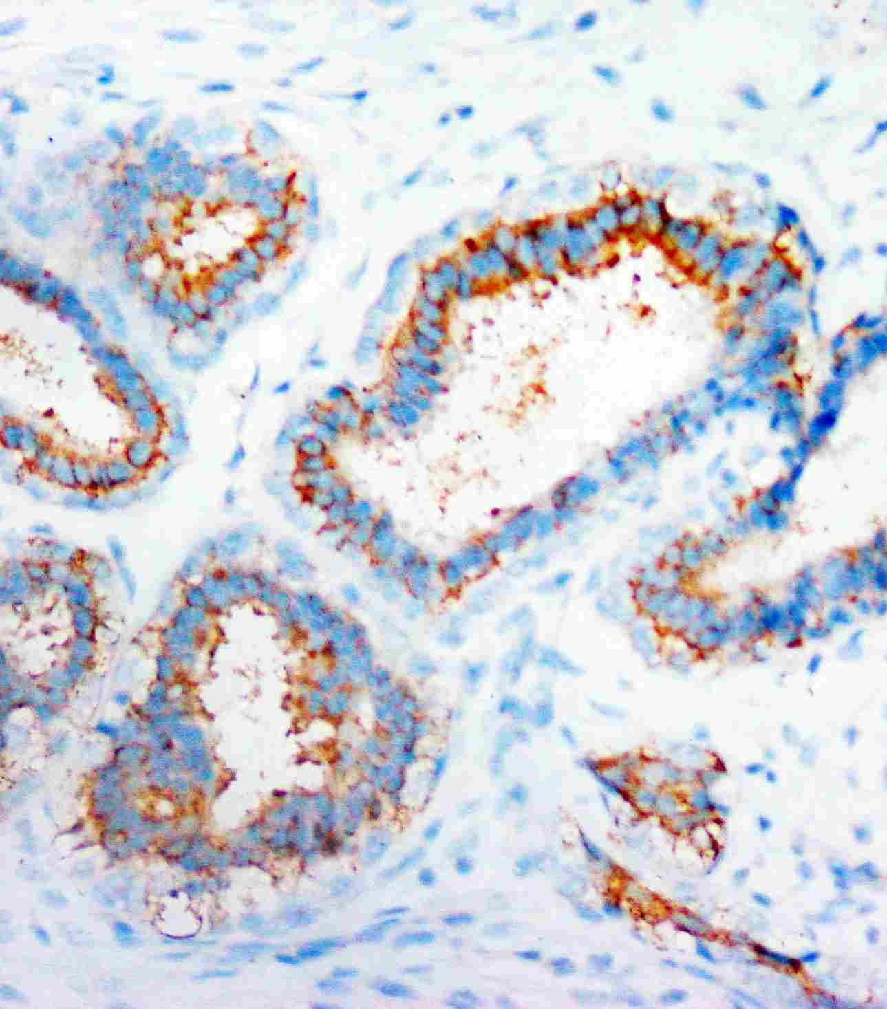
- 产品详情
- 实验流程
- 背景知识
Application 
| WB, IHC-P |
|---|---|
| Primary Accession | P61073 |
| Host | Rabbit |
| Reactivity | Human, Mouse, Rat |
| Clonality | Polyclonal |
| Format | Lyophilized |
| Description | Rabbit IgG polyclonal antibody for C-X-C chemokine receptor type 4(CXCR4) detection. Tested with WB, IHC-P in Human;Mouse;Rat. |
| Reconstitution | Add 0.2ml of distilled water will yield a concentration of 500ug/ml. |
| Gene ID | 7852 |
|---|---|
| Other Names | C-X-C chemokine receptor type 4, CXC-R4, CXCR-4, FB22, Fusin, HM89, LCR1, Leukocyte-derived seven transmembrane domain receptor, LESTR, Lipopolysaccharide-associated protein 3, LAP-3, LPS-associated protein 3, NPYRL, Stromal cell-derived factor 1 receptor, SDF-1 receptor, CD184, CXCR4 |
| Calculated MW | 39746 Da |
| Application Details | Immunohistochemistry(Paraffin-embedded Section), 0.5-1 µg/ml, Human, By Heat Western blot, 0.1-0.5 µg/ml, Human, Rat, Mouse |
| Subcellular Localization | Cell membrane; Multi-pass membrane protein. Cell junction. Early endosome. Late endosome. Lysosome. In unstimulated cells, diffuse pattern on plasma membrane. On agonist stimulation, colocalizes with ITCH at the plasma membrane where it becomes ubiquitinated. In the presence of antigen, distributes to the immunological synapse forming at the T-cell-APC contact area, where it localizes at the peripheral and distal supramolecular activation cluster (SMAC). |
| Tissue Specificity | Expressed in numerous tissues, such as peripheral blood leukocytes, spleen, thymus, spinal cord, heart, placenta, lung, liver, skeletal muscle, kidney, pancreas, cerebellum, cerebral cortex and medulla (in microglia as well as in astrocytes), brain microvascular, coronary artery and umbilical cord endothelial cells. Isoform 1 is predominant in all tissues tested. . |
| Source | Eukaryota |
| Protein Name | C-X-C chemokine receptor type 4(CXC-R4/CXCR-4) |
| Contents | Each vial contains 5mg BSA, 0.9mg NaCl, 0.2mg Na2HPO4, 0.05mg Thimerosal, 0.05mg NaN3. |
| Immunogen | A synthetic peptide corresponding to a sequence at the N-terminus of human CXCR4(18-33aa SGDYDSMKEPCFREEN), different from the related rat and mouse sequences by two amino acids. |
| Purification | Immunogen affinity purified. |
| Cross Reactivity | No cross reactivity with other proteins |
| Storage | At -20˚C for one year. After r˚Constitution, at 4˚C for one month. It˚Can also be aliquotted and stored frozen at -20˚C for a longer time.Avoid repeated freezing and thawing. |
| Sequence Similarities | Belongs to the G-protein coupled receptor 1 family. |
For Research Use Only. Not For Use In Diagnostic Procedures.
| Name | CXCR4 {ECO:0000303|PubMed:9468539, ECO:0000312|HGNC:HGNC:2561} |
|---|---|
| Function | Receptor for the C-X-C chemokine CXCL12/SDF-1 that transduces a signal by increasing intracellular calcium ion levels and enhancing MAPK1/MAPK3 activation (PubMed:10074102, PubMed:10452968, PubMed:10644702, PubMed:10825158, PubMed:18799424, PubMed:20048153, PubMed:20505072, PubMed:24912431, PubMed:28978524, PubMed:8752280, PubMed:8752281). Ligand binding causes a conformation change that triggers signaling via guanine nucleotide-binding proteins (G proteins) and modulates the activity of downstream effectors, such as adenylate cyclase (PubMed:16725153, PubMed:17197449, PubMed:18799424, PubMed:39093700). CXCR4 is coupled to G(i) G alpha proteins and mediates inhibition of adenylate cyclase (PubMed:17197449, PubMed:39093700). Involved in the AKT signaling cascade (PubMed:24912431). Plays a role in regulation of cell migration, e.g. during wound healing (PubMed:28978524). Also acts as a receptor for extracellular ubiquitin; leading to enhanced intracellular calcium ions and reduced cellular cAMP levels (PubMed:20228059). Binds bacterial lipopolysaccharide (LPS) et mediates LPS-induced inflammatory response, including TNF secretion by monocytes (PubMed:11276205). Involved in hematopoiesis and in cardiac ventricular septum formation (By similarity). Also plays an essential role in vascularization of the gastrointestinal tract, probably by regulating vascular branching and/or remodeling processes in endothelial cells (By similarity). Involved in cerebellar development; in the CNS, could mediate hippocampal-neuron surviva (By similarity). |
| Cellular Location | Cell membrane; Multi-pass membrane protein. Cell junction. Early endosome. Late endosome. Lysosome Note=In unstimulated cells, diffuse pattern on plasma membrane (PubMed:10452968, PubMed:14602072, PubMed:21540189). On agonist stimulation, colocalizes with ITCH at the plasma membrane where it becomes ubiquitinated (PubMed:14602072). In the presence of antigen, distributes to the immunological synapse forming at the T-cell-APC contact area, where it localizes at the peripheral and distal supramolecular activation cluster (SMAC) (PubMed:20215400) |
| Tissue Location | Expressed in numerous tissues, such as peripheral blood leukocytes, spleen, thymus, spinal cord, heart, placenta, lung, liver, skeletal muscle, kidney, pancreas, cerebellum, cerebral cortex and medulla (in microglia as well as in astrocytes), brain microvascular, coronary artery and umbilical cord endothelial cells Isoform 1 is predominant in all tissues tested |
Provided below are standard protocols that you may find useful for product applications.
BACKGROUND
CXCR4 (Chemokine,CXC Motif, Receptor 4), also known as FUSIN or NPY3R, is a protein that in humans is encoded by the CXCR4 gene. It is the receptor for the CXC chemokine SDF1 that has essential functions on embryo organogenesis, immunological functions and T lymphocyte trafficking. CXCR4 is the only SDF1 receptor identified so far. This suggests that CXCR4 expression is critical for the biological effects of SDF1. CXCR4 is also a seven-transmembrane-spanning, G-protein-coupled receptor for the CXC chemokine PBSF/SDF-1. It functions as a co-receptor for T-cell-line tropic human immunodeficiency virus HIV-1. It was concluded that PBSF/SDF-1 and CXCR4 define a new signalling system for organ vascularization.

终于等到您。ABCEPTA(百远生物)抗体产品。
点击下方“我要评价 ”按钮提交您的反馈信息,您的反馈和评价是我们最宝贵的财富之一,
我们将在1-3个工作日内处理您的反馈信息。
如有疑问,联系:0512-88856768 tech-china@abcepta.com.


