


 癌症的基本特征包括细胞增殖、血管生成、迁移、凋亡逃避机制和细胞永生等。找到癌症发生过程中这些通路的关键标记物和对应的抗体用于检测至关重要。
 为您推荐一个泛素化位点预测神器——泛素化分析工具,可以为您的蛋白的泛素化位点作出预测和评分。
 细胞自噬受体图形绘图工具为你的蛋白的细胞受体结合位点作出预测和评分,识别结合到自噬通路中的蛋白是非常重要的,便于让我们理解自噬在正常生理、病理过程中的作用,如发育、细胞分化、神经退化性疾病、压力条件下、感染和癌症。
Kl antibody - N-terminal region
Rabbit Polyclonal Antibody
- 产品详情
- 实验流程
Application
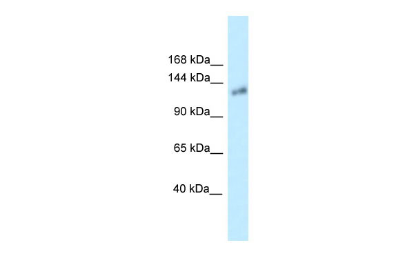
| WB |
|---|---|
| Primary Accession | O35082 |
| Other Accession | NM_013823, NP_038851 |
| Reactivity | Human, Mouse, Rat, Rabbit, Pig, Horse, Bovine |
| Predicted | Human, Mouse, Rat, Rabbit, Pig, Chicken, Bovine |
| Host | Rabbit |
| Clonality | Polyclonal |
| Calculated MW | 116398 Da |
| Gene ID | 16591 |
|---|---|
| Alias Symbol | alpha-kl |
| Other Names | Klotho, 3.2.1.31, Klotho peptide, Kl |
| Format | Liquid. Purified antibody supplied in 1x PBS buffer with 0.09% (w/v) sodium azide and 2% sucrose. |
| Reconstitution & Storage | Add 50 ul of distilled water. Final anti-Kl antibody concentration is 1 mg/ml in PBS buffer with 2% sucrose. For longer periods of storage, store at 20°C. Avoid repeat freeze-thaw cycles. |
| Precautions | Kl antibody - N-terminal region is for research use only and not for use in diagnostic or therapeutic procedures. |
| Name | Kl |
|---|---|
| Function | May have weak glycosidase activity towards glucuronylated steroids. However, it lacks essential active site Glu residues at positions 241 and 874, suggesting it may be inactive as a glycosidase in vivo. May be involved in the regulation of calcium and phosphorus homeostasis by inhibiting the synthesis of active vitamin D. Essential factor for the specific interaction between FGF23 and FGFR1. |
| Cellular Location | [Isoform 1]: Cell membrane; Single-pass type I membrane protein. Apical cell membrane; Single-pass type I membrane protein. Note=Isoform 1 shedding leads to a soluble peptide. [Klotho peptide]: Secreted |
| Tissue Location | Membrane-bound protein is present in distal renal tubules, inner ear, ependymal cells of brain choroid plexus, elongating spermatids and mature oocytes (at protein level). Soluble peptide is present in serum (100 pM) and cerebrospinal fluid. Expressed strongly in kidney, moderately in brain choroid plexus, and at low levels in pituitary, placenta, skeletal muscle, urinary bladder, aorta, pancreas, testis, ovary, colon, thyroid gland and adipocytes |
Research Areas
For Research Use Only. Not For Use In Diagnostic Procedures.
Application Protocols
Provided below are standard protocols that you may find useful for product applications.
终于等到您。ABCEPTA(百远生物)抗体产品。
点击下方“我要评价 ”按钮提交您的反馈信息,您的反馈和评价是我们最宝贵的财富之一,
我们将在1-3个工作日内处理您的反馈信息。
如有疑问,联系:0512-88856768 tech-china@abcepta.com.


