


 癌症的基本特征包括细胞增殖、血管生成、迁移、凋亡逃避机制和细胞永生等。找到癌症发生过程中这些通路的关键标记物和对应的抗体用于检测至关重要。
 为您推荐一个泛素化位点预测神器——泛素化分析工具,可以为您的蛋白的泛素化位点作出预测和评分。
 细胞自噬受体图形绘图工具为你的蛋白的细胞受体结合位点作出预测和评分,识别结合到自噬通路中的蛋白是非常重要的,便于让我们理解自噬在正常生理、病理过程中的作用,如发育、细胞分化、神经退化性疾病、压力条件下、感染和癌症。
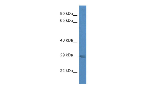
Rab23 antibody - middle region
Rabbit Polyclonal Antibody
- 产品详情
- 实验流程
Application
| WB |
|---|---|
| Primary Accession | P35288 |
| Other Accession | NM_008999, NP_033025 |
| Reactivity | Human, Mouse, Rat, Rabbit, Zebrafish, Pig, Dog, Guinea Pig, Horse, Bovine |
| Predicted | Human, Mouse, Rat, Zebrafish, Chicken, Dog, Bovine |
| Host | Rabbit |
| Clonality | Polyclonal |
| Calculated MW | 26678 Da |
| Alias Symbol | AW545388, opb, opb2 |
|---|---|
| Other Names | Ras-related protein Rab-23, Protein open brain, Rab-15, Rab23, Opb |
| Format | Liquid. Purified antibody supplied in 1x PBS buffer with 0.09% (w/v) sodium azide and 2% sucrose. |
| Reconstitution & Storage | Add 50 ul of distilled water. Final anti-Rab23 antibody concentration is 1 mg/ml in PBS buffer with 2% sucrose. For longer periods of storage, store at 20°C. Avoid repeat freeze-thaw cycles. |
| Precautions | Rab23 antibody - middle region is for research use only and not for use in diagnostic or therapeutic procedures. |
| Name | Rab23 {ECO:0000312|MGI:MGI:99833} |
|---|---|
| Function | The small GTPases Rab are key regulators of intracellular membrane trafficking, from the formation of transport vesicles to their fusion with membranes. Rabs cycle between an inactive GDP-bound form and an active GTP-bound form that is able to recruit to membranes different set of downstream effectors directly responsible for vesicle formation, movement, tethering and fusion (By similarity). In conjunction with IFT57 and KIF17, it drives the localization of specific G protein-coupled receptors, such as the dopamime receptor DRD1, to primary cilia (By similarity). Has a critical role in the formation and elongation of neuronal primary cilia, thereby impacting the activation of sonic hedgehog (Shh) signaling (PubMed:40825043). Additionally, it is involved in the down-regulation of Shh signaling by cooperating with SUFU to prevent the nuclear import of GLI1 transcription factor, thus suppressing its transcriptional activity (PubMed:11449277, PubMed:16364285). Regulates GLI1 in differentiating chondrocytes. Likewise, regulates GLI3 proteolytic processing and modulates GLI2 and GLI3 transcription factor activity. Plays a role in autophagic vacuole assembly, and mediates defense against pathogens, such as S.aureus, by promoting their capture by autophagosomes that then merge with lysosomes (By similarity). |
| Cellular Location | Cell membrane; Lipid-anchor; Cytoplasmic side. Cytoplasm Endosome membrane. Cytoplasmic vesicle, autophagosome {ECO:0000250|UniProtKB:Q9ULC3}. Cytoplasmic vesicle, phagosome. Cytoplasmic vesicle, phagosome membrane; Lipid-anchor; Cytoplasmic side. Note=Recruited to phagosomes containing S.aureus or Mycobacterium. {ECO:0000250|UniProtKB:Q9ULC3} |
| Tissue Location | Detected in brain neurons (at protein level). Forebrain and midbrain. |
Research Areas
For Research Use Only. Not For Use In Diagnostic Procedures.
Application Protocols
Provided below are standard protocols that you may find useful for product applications.
REFERENCES
Olkkonen V.M.,et al.Gene 138:207-211(1994).
Chavrier P.,et al.Gene 112:261-264(1992).
Gunther T.,et al.Development 120:3119-3130(1994).
Eggenschwiler J.T.,et al.Dev. Biol. 227:648-660(2000).
Eggenschwiler J.T.,et al.Nature 412:194-198(2001).
终于等到您。ABCEPTA(百远生物)抗体产品。
点击下方“我要评价 ”按钮提交您的反馈信息,您的反馈和评价是我们最宝贵的财富之一,
我们将在1-3个工作日内处理您的反馈信息。
如有疑问,联系:0512-88856768 tech-china@abcepta.com.


