


 癌症的基本特征包括细胞增殖、血管生成、迁移、凋亡逃避机制和细胞永生等。找到癌症发生过程中这些通路的关键标记物和对应的抗体用于检测至关重要。
 为您推荐一个泛素化位点预测神器——泛素化分析工具,可以为您的蛋白的泛素化位点作出预测和评分。
 细胞自噬受体图形绘图工具为你的蛋白的细胞受体结合位点作出预测和评分,识别结合到自噬通路中的蛋白是非常重要的,便于让我们理解自噬在正常生理、病理过程中的作用,如发育、细胞分化、神经退化性疾病、压力条件下、感染和癌症。
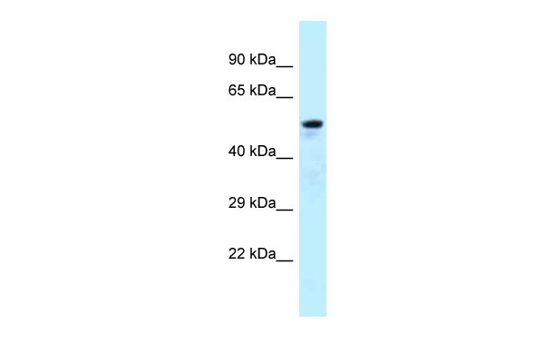
ARFGAP3 antibody - C-terminal region
Rabbit Polyclonal Antibody
- 产品详情
- 实验流程
Application 
| WB |
|---|---|
| Primary Accession | Q9NP61 |
| Other Accession | NM_014570, NP_055385 |
| Reactivity | Human, Rat, Pig, Dog, Horse, Bovine |
| Predicted | Human, Dog, Horse |
| Host | Rabbit |
| Clonality | Polyclonal |
| Calculated MW | 56928 Da |
| Gene ID | 26286 |
|---|---|
| Alias Symbol | ARFGAP1, FLJ45618 |
| Other Names | ADP-ribosylation factor GTPase-activating protein 3, ARF GAP 3, ARFGAP3, ARFGAP1 |
| Format | Liquid. Purified antibody supplied in 1x PBS buffer with 0.09% (w/v) sodium azide and 2% sucrose. |
| Reconstitution & Storage | Add 50 ul of distilled water. Final anti-ARFGAP3 antibody concentration is 1 mg/ml in PBS buffer with 2% sucrose. For longer periods of storage, store at 20°C. Avoid repeat freeze-thaw cycles. |
| Precautions | ARFGAP3 antibody - C-terminal region is for research use only and not for use in diagnostic or therapeutic procedures. |
For Research Use Only. Not For Use In Diagnostic Procedures.
| Name | ARFGAP3 |
|---|---|
| Synonyms | ARFGAP1 |
| Function | GTPase-activating protein (GAP) for ADP ribosylation factor 1 (ARF1), which is essential for COPI vesicle formation and retrograde transport between the Golgi and the endoplasmic reticulum (PubMed:11172815, PubMed:20858901). Hydrolysis of ARF1-bound GTP leads to dissociation of coatomer from Golgi-derived membranes to allow fusion with target membranes (PubMed:11172815, PubMed:20858901). Promotes the proper assembly of the COPI coat lattice, rather than just acting as a simple curvature-dependent GAP like ARFGAP1 (PubMed:20858901). |
| Cellular Location | Golgi apparatus membrane; Peripheral membrane protein; Cytoplasmic side. Cytoplasm Note=Also found on peripheral punctate structures likely to be endoplasmic reticulum-Golgi intermediate compartment |
| Tissue Location | Widely expressed. Highest expression in endocrine glands (pancreas, pituitary gland, salivary gland, and prostate) and testis with a much higher expression in the testis than in the ovary |
Research Areas
Application Protocols
Provided below are standard protocols that you may find useful for product applications.
REFERENCES
Zhang C.,et al.Genomics 63:400-408(2000).
Collins J.E.,et al.Genome Res. 13:27-36(2003).
Collins J.E.,et al.Genome Biol. 5:R84.1-R84.11(2004).
Dunham I.,et al.Nature 402:489-495(1999).
Bechtel S.,et al.BMC Genomics 8:399-399(2007).

终于等到您。ABCEPTA(百远生物)抗体产品。
点击下方“我要评价 ”按钮提交您的反馈信息,您的反馈和评价是我们最宝贵的财富之一,
我们将在1-3个工作日内处理您的反馈信息。
如有疑问,联系:0512-88856768 tech-china@abcepta.com.


