


 癌症的基本特征包括细胞增殖、血管生成、迁移、凋亡逃避机制和细胞永生等。找到癌症发生过程中这些通路的关键标记物和对应的抗体用于检测至关重要。
 为您推荐一个泛素化位点预测神器——泛素化分析工具,可以为您的蛋白的泛素化位点作出预测和评分。
 细胞自噬受体图形绘图工具为你的蛋白的细胞受体结合位点作出预测和评分,识别结合到自噬通路中的蛋白是非常重要的,便于让我们理解自噬在正常生理、病理过程中的作用,如发育、细胞分化、神经退化性疾病、压力条件下、感染和癌症。
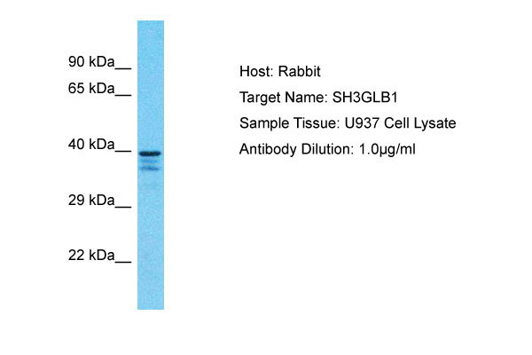
SH3GLB1 Antibody - C-terminal region
Rabbit Polyclonal Antibody
- 产品详情
- 实验流程
- 背景知识
Application
| WB |
|---|---|
| Primary Accession | Q9Y371 |
| Other Accession | NP_057093 |
| Reactivity | Human |
| Host | Rabbit |
| Clonality | Polyclonal |
| Calculated MW | 40796 Da |
| Gene ID | 51100 |
|---|---|
| Alias Symbol | SH3GLB1, KIAA0491, CGI-61, |
| Other Names | Endophilin-B1, Bax-interacting factor 1, Bif-1, SH3 domain-containing GRB2-like protein B1, SH3GLB1, KIAA0491 |
| Format | Liquid. Purified antibody supplied in 1x PBS buffer with 0.09% (w/v) sodium azide and 2% sucrose. |
| Reconstitution & Storage | Add 50 &mu, l of distilled water. Final Anti-SH3GLB1 antibody concentration is 1 mg/ml in PBS buffer with 2% sucrose. For longer periods of storage, store at -20°C. Avoid repeat freeze-thaw cycles. |
| Precautions | SH3GLB1 Antibody - C-terminal region is for research use only and not for use in diagnostic or therapeutic procedures. |
| Name | SH3GLB1 |
|---|---|
| Synonyms | KIAA0491 |
| Function | May be required for normal outer mitochondrial membrane dynamics (PubMed:15452144). Required for coatomer-mediated retrograde transport in certain cells (By similarity). May recruit other proteins to membranes with high curvature. May promote membrane fusion (PubMed:11604418). Involved in activation of caspase-dependent apoptosis by promoting BAX/BAK1 activation (PubMed:16227588). Isoform 1 acts proapoptotic in fibroblasts (By similarity). Involved in caspase- independent apoptosis during nutrition starvation and involved in the regulation of autophagy. Activates lipid kinase activity of PIK3C3 during autophagy probably by associating with the PI3K complex II (PI3KC3-C2) (PubMed:17891140). Associated with PI3KC3-C2 during autophagy may regulate the trafficking of ATG9A from the Golgi complex to the peripheral cytoplasm for the formation of autophagosomes by inducing Golgi membrane tubulation and fragmentation (PubMed:21068542). Involved in regulation of degradative endocytic trafficking and cytokinesis, probably in the context of PI3KC3-C2 (PubMed:20643123). Isoform 2 acts antiapoptotic in neuronal cells; involved in maintenance of mitochondrial morphology and promotes neuronal viability (By similarity). |
| Cellular Location | Cytoplasm. Golgi apparatus membrane; Peripheral membrane protein. Mitochondrion outer membrane; Peripheral membrane protein. Cytoplasmic vesicle, autophagosome membrane. Midbody. Note=Association with the Golgi apparatus depends on the cell type (By similarity). Following starvation colocalizes with ATG5 and LC3 autophagy-related protein(s)on autophagosomal membranes (PubMed:17891140). {ECO:0000250, ECO:0000269|PubMed:17891140} |
| Tissue Location | Highly expressed in heart, skeletal muscle, kidney and placenta. Detected at lower levels in brain, colon, thymus, spleen, liver, small intestine, lung and peripheral blood leukocytes |
For Research Use Only. Not For Use In Diagnostic Procedures.
Provided below are standard protocols that you may find useful for product applications.
BACKGROUND
May be required for normal outer mitochondrial membrane dynamics. Required for coatomer-mediated retrograde transport in certain cells. May recruit other proteins to membranes with high curvature. May promote membrane fusion.
REFERENCES
Pierrat B.,et al.Genomics 71:222-234(2001).
Cuddeback S.M.,et al.J. Biol. Chem. 276:20559-20565(2001).
Modregger J.,et al.J. Biol. Chem. 278:4160-4167(2003).
Seki N.,et al.DNA Res. 4:345-349(1997).
Lai C.-H.,et al.Genome Res. 10:703-713(2000).
终于等到您。ABCEPTA(百远生物)抗体产品。
点击下方“我要评价 ”按钮提交您的反馈信息,您的反馈和评价是我们最宝贵的财富之一,
我们将在1-3个工作日内处理您的反馈信息。
如有疑问,联系:0512-88856768 tech-china@abcepta.com.


