


 癌症的基本特征包括细胞增殖、血管生成、迁移、凋亡逃避机制和细胞永生等。找到癌症发生过程中这些通路的关键标记物和对应的抗体用于检测至关重要。
 为您推荐一个泛素化位点预测神器——泛素化分析工具,可以为您的蛋白的泛素化位点作出预测和评分。
 细胞自噬受体图形绘图工具为你的蛋白的细胞受体结合位点作出预测和评分,识别结合到自噬通路中的蛋白是非常重要的,便于让我们理解自噬在正常生理、病理过程中的作用,如发育、细胞分化、神经退化性疾病、压力条件下、感染和癌症。
PINK1 Antibody (Ascites)
Unpurified Mouse Monoclonal Antibody (Mab)
- 产品详情
- 文献引用 : 3
- 实验流程
- 背景知识
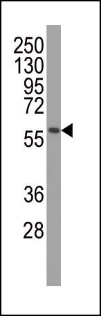
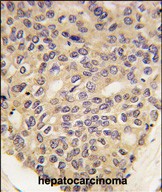
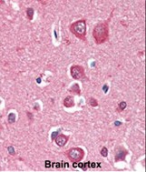
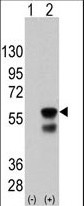
Application
| WB, IHC-P, E |
|---|---|
| Primary Accession | Q9BXM7 |
| Reactivity | Human, Mouse |
| Host | Mouse |
| Clonality | Monoclonal |
| Isotype | IgG1 |
| Clone Names | 38CT20.8.5 |
| Calculated MW | 62769 Da |
| Gene ID | 65018 |
|---|---|
| Other Names | Serine/threonine-protein kinase PINK1, mitochondrial, BRPK, PTEN-induced putative kinase protein 1, PINK1 |
| Target/Specificity | Recombinant PINK1 protein was used to produced this monoclonal antibody. |
| Dilution | WB~~1:500~2000 IHC-P~~1:100~500 E~~Use at an assay dependent concentration. |
| Format | Mouse monoclonal antibody supplied in crude ascites with 0.09% (W/V) sodium azide. |
| Storage | Maintain refrigerated at 2-8°C for up to 2 weeks. For long term storage store at -20°C in small aliquots to prevent freeze-thaw cycles. |
| Precautions | PINK1 Antibody (Ascites) is for research use only and not for use in diagnostic or therapeutic procedures. |
| Name | PINK1 |
|---|---|
| Function | Serine/threonine-protein kinase which acts as a sensor of mitochondrial damage and protects against mitochondrial dysfunction during cellular stress (PubMed:40080546). It phosphorylates mitochondrial proteins to coordinate mitochondrial quality control mechanisms that remove and replace dysfunctional mitochondrial components (PubMed:14607334, PubMed:15087508, PubMed:18443288, PubMed:18957282, PubMed:19229105, PubMed:19966284, PubMed:20404107, PubMed:20547144, PubMed:20798600, PubMed:22396657, PubMed:23620051, PubMed:23754282, PubMed:23933751, PubMed:24660806, PubMed:24751536, PubMed:24784582, PubMed:24896179, PubMed:24898855, PubMed:25527291, PubMed:32484300). In healthy mitochondria, PINK1 is translocated across the mitochondrial outer membrane (MOM) via the translocase of the outer membrane (TOM) complex, and inserted into the mitochondrial inner membrane (MIM) via the translocase of the inner membrane (TIM23) complex where it is cleaved and released into the cytosol (PubMed:40080546). Depending on the severity of mitochondrial damage, activity ranges from preventing apoptosis and stimulating mitochondrial biogenesis to eliminating severely damaged mitochondria via PINK1-PRKN- dependent mitophagy (PubMed:14607334, PubMed:15087508, PubMed:18443288, PubMed:19966284, PubMed:20404107, PubMed:20798600, PubMed:22396657, PubMed:23620051, PubMed:23933751, PubMed:24898855, PubMed:32047033, PubMed:32484300). When cellular stress results in irreversible mitochondrial damage, PINK1 accumulates at the outer mitochondrial membrane (OMM) where it phosphorylates pre-existing polyubiquitin chains at 'Ser-65', recruits PRKN from the cytosol to the OMM and activates PRKN by phosphorylation at 'Ser-65'; activated PRKN then ubiquitinates VDAC1 and other OMM proteins to initiate mitophagy (PubMed:14607334, PubMed:15087508, PubMed:19966284, PubMed:20404107, PubMed:20798600, PubMed:23754282, PubMed:23933751, PubMed:24660806, PubMed:24751536, PubMed:24784582, PubMed:25474007, PubMed:25527291, PubMed:32047033, PubMed:40080546). The PINK1-PRKN pathway also promotes fission of damaged mitochondria through phosphorylation and PRKN- dependent degradation of mitochondrial proteins involved in fission such as MFN2 (PubMed:18443288, PubMed:23620051, PubMed:24898855). This prevents the refusion of unhealthy mitochondria with the mitochondrial network or initiates mitochondrial fragmentation facilitating their later engulfment by autophagosomes (PubMed:18443288, PubMed:23620051). Also promotes mitochondrial fission independently of PRKN and ATG7- mediated mitophagy, via the phosphorylation and activation of DNM1L (PubMed:18443288, PubMed:32484300). Regulates motility of damaged mitochondria by promoting the ubiquitination and subsequent degradation of MIRO1 and MIRO2; in motor neurons, this likely inhibits mitochondrial intracellular anterograde transport along the axons which probably increases the chance of the mitochondria undergoing mitophagy in the soma (PubMed:22396657). Required for ubiquinone reduction by mitochondrial complex I by mediating phosphorylation of complex I subunit NDUFA10 (By similarity). Phosphorylates LETM1, positively regulating its mitochondrial calcium transport activity (PubMed:29123128). |
| Cellular Location | Mitochondrion outer membrane Mitochondrion inner membrane {ECO:0000250|UniProtKB:Q99MQ3}. Cytoplasm, cytosol Note=Localizes mostly in mitochondrion and the two smaller proteolytic processed fragments localize mainly in cytosol (PubMed:19229105). In healthy mitochondria, PINK1 is translocated across the mitochondrial membranes (PubMed:40080546). Upon mitochondrial membrane depolarization following damage, PINK1 import is stalled, which induces its accumulation in the outer mitochondrial membrane and the activation of its kinase activity (PubMed:18957282, PubMed:40080546) |
| Tissue Location | Highly expressed in heart, skeletal muscle and testis, and at lower levels in brain, placenta, liver, kidney, pancreas, prostate, ovary and small intestine. Present in the embryonic testis from an early stage of development |
For Research Use Only. Not For Use In Diagnostic Procedures.

Provided below are standard protocols that you may find useful for product applications.
BACKGROUND
This gene encodes a serine/threonine protein kinase that localizes to mitochondria. It is thought to protect cells from stress-induced mitochondrial dysfunction. Mutations in this gene cause one form of autosomal recessive early-onset Parkinson disease.
REFERENCES
Oxidative stress alters the regulatory control of p66Shc and Akt in PINK1 deficient cells. Maj MC, et al. Biochem Biophys Res Commun, 2010 Aug 27. PMID 20637729. Assessing the prevalence of PINK1 genetic variants in South African patients diagnosed with early- and late-onset Parkinson's disease. Keyser RJ, et al. Biochem Biophys Res Commun, 2010 Jul 16. PMID 20558144. Progression of subtle motor signs in PINK1 mutation carriers with mild dopaminergic deficit. Eggers C, et al. Neurology, 2010 Jun 1. PMID 20513816. Structural imaging in the presymptomatic stage of genetically determined parkinsonism. Reetz K, et al. Neurobiol Dis, 2010 Sep. PMID 20483373. Clinical and demographic characteristics of PINK1 mutation carriers--a meta-analysis. Kasten M, et al. Mov Disord, 2010 May 15. PMID 20461815.
终于等到您。ABCEPTA(百远生物)抗体产品。
点击下方“我要评价 ”按钮提交您的反馈信息,您的反馈和评价是我们最宝贵的财富之一,
我们将在1-3个工作日内处理您的反馈信息。
如有疑问,联系:0512-88856768 tech-china@abcepta.com.


