


 癌症的基本特征包括细胞增殖、血管生成、迁移、凋亡逃避机制和细胞永生等。找到癌症发生过程中这些通路的关键标记物和对应的抗体用于检测至关重要。
 为您推荐一个泛素化位点预测神器——泛素化分析工具,可以为您的蛋白的泛素化位点作出预测和评分。
 细胞自噬受体图形绘图工具为你的蛋白的细胞受体结合位点作出预测和评分,识别结合到自噬通路中的蛋白是非常重要的,便于让我们理解自噬在正常生理、病理过程中的作用,如发育、细胞分化、神经退化性疾病、压力条件下、感染和癌症。
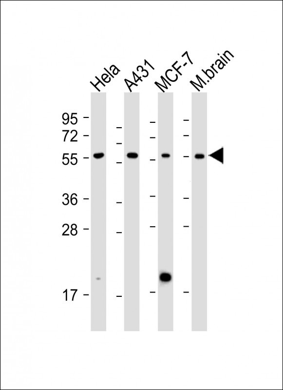
PINK1 Antibody
Purified Mouse Monoclonal Antibody (Mab)
- 产品详情
- 实验流程
- 背景知识
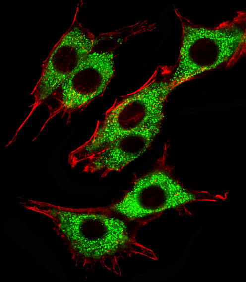
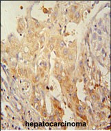
Application
| WB, IHC-P, IF, E |
|---|---|
| Primary Accession | Q9BXM7 |
| Reactivity | Human, Mouse |
| Host | Mouse |
| Clonality | Monoclonal |
| Isotype | IgG1 |
| Clone Names | 38CT18.2.6 |
| Antigen Region | Unknown aa |
| Other Names | Serine/threonine-protein kinase PINK1, mitochondrial, BRPK, PTEN-induced putative kinase protein 1, PINK1 |
|---|---|
| Target/Specificity | Recombinant PINK1 protein was used to produced this monoclonal antibody. |
| Dilution | WB~~1:4000 IHC-P~~1:100~500 IF~~1:25 E~~Use at an assay dependent concentration. |
| Format | Purified monoclonal antibody supplied in PBS with 0.09% (W/V) sodium azide. This antibody is purified through a protein G column, followed by dialysis against PBS. |
| Storage | Maintain refrigerated at 2-8°C for up to 2 weeks. For long term storage store at -20°C in small aliquots to prevent freeze-thaw cycles. |
| Precautions | PINK1 Antibody is for research use only and not for use in diagnostic or therapeutic procedures. |
For Research Use Only. Not For Use In Diagnostic Procedures.
Provided below are standard protocols that you may find useful for product applications.
BACKGROUND
This gene encodes a serine/threonine protein kinase that localizes to mitochondria. It is thought to protect cells from stress-induced mitochondrial dysfunction. Mutations in this gene cause one form of autosomal recessive early-onset Parkinson disease.
REFERENCES
Oxidative stress alters the regulatory control of p66Shc and Akt in PINK1 deficient cells. Maj MC, et al. Biochem Biophys Res Commun, 2010 Aug 27. PMID 20637729.
Assessing the prevalence of PINK1 genetic variants in South African patients diagnosed with early- and late-onset Parkinson's disease. Keyser RJ, et al. Biochem Biophys Res Commun, 2010 Jul 16. PMID 20558144.
Progression of subtle motor signs in PINK1 mutation carriers with mild dopaminergic deficit. Eggers C, et al. Neurology, 2010 Jun 1. PMID 20513816.
Structural imaging in the presymptomatic stage of genetically determined parkinsonism. Reetz K, et al. Neurobiol Dis, 2010 Sep. PMID 20483373.
Clinical and demographic characteristics of PINK1 mutation carriers--a meta-analysis. Kasten M, et al. Mov Disord, 2010 May 15. PMID 20461815.
终于等到您。ABCEPTA(百远生物)抗体产品。
点击下方“我要评价 ”按钮提交您的反馈信息,您的反馈和评价是我们最宝贵的财富之一,
我们将在1-3个工作日内处理您的反馈信息。
如有疑问,联系:0512-88856768 tech-china@abcepta.com.


