


 癌症的基本特征包括细胞增殖、血管生成、迁移、凋亡逃避机制和细胞永生等。找到癌症发生过程中这些通路的关键标记物和对应的抗体用于检测至关重要。
 为您推荐一个泛素化位点预测神器——泛素化分析工具,可以为您的蛋白的泛素化位点作出预测和评分。
 细胞自噬受体图形绘图工具为你的蛋白的细胞受体结合位点作出预测和评分,识别结合到自噬通路中的蛋白是非常重要的,便于让我们理解自噬在正常生理、病理过程中的作用,如发育、细胞分化、神经退化性疾病、压力条件下、感染和癌症。
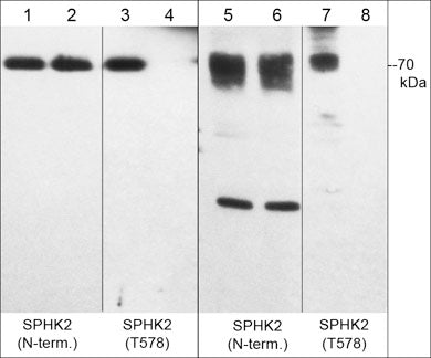
Anti-Sphingosine Kinase 2 (N-terminal region) Antibody
- 产品详情
- 实验流程
- 背景知识
Application
| WB, ICC, IP |
|---|---|
| Primary Accession | Q9NRA0 |
| Host | Rabbit |
| Clonality | Rabbit Polyclonal |
| Isotype | IgG |
| Calculated MW | 69217 Da |
| Gene ID | 56848 |
|---|---|
| Other Names | SK2, Spk2, Sphk2 |
| Dilution | WB~~1:1000 ICC~~N/A IP~~N/A |
| Storage | Maintain refrigerated at 2-8°C for up to 6 months. For long term storage store at -20°C in small aliquots to prevent freeze-thaw cycles. |
| Precautions | Anti-Sphingosine Kinase 2 (N-terminal region) Antibody is for research use only and not for use in diagnostic or therapeutic procedures. |
| Shipping | Blue Ice |
For Research Use Only. Not For Use In Diagnostic Procedures.
Provided below are standard protocols that you may find useful for product applications.
BACKGROUND
Sphingolipids are metabolized into bioactive products that include ceramide, sphingosine, and sphingosine-1-phosphate (S1P). Sphingosine Kinase (SK) catalyzes the phosphorylation of the lipid sphingosine, creating S1P. S1P subsequently signals through cell surface G protein-coupled receptors, as well as intracellularly, to modulate cell proliferation, survival, motility and differentiation. Two isoforms of SK have been identified, SK1 and SK2. The mRNA for both of these isoforms is widely expressed with SK1 expression highest in brain, heart, kidney, thymus, spleen and lung, while SK2 is highest in kidney and liver. SKs can be activated through growth factor, G protein-coupled, and immunoglobulin receptor signalling. Regulation of SK1 and SK2 activity may occur through phosphorylation. SK1 is phosphorylated at Ser-225 by ERK leading to increased activity and translocation to the plasma membrane. SK2 is phosphorylated in response to EGF, PKC activators, and phorbol esters. ERK1 can phosphorylate both Ser-351 and Thr-578, and non-phosphorylatable mutants of these sites suppress ERK1-mediated chemotaxis.
终于等到您。ABCEPTA(百远生物)抗体产品。
点击下方“我要评价 ”按钮提交您的反馈信息,您的反馈和评价是我们最宝贵的财富之一,
我们将在1-3个工作日内处理您的反馈信息。
如有疑问,联系:0512-88856768 tech-china@abcepta.com.


