


 癌症的基本特征包括细胞增殖、血管生成、迁移、凋亡逃避机制和细胞永生等。找到癌症发生过程中这些通路的关键标记物和对应的抗体用于检测至关重要。
 为您推荐一个泛素化位点预测神器——泛素化分析工具,可以为您的蛋白的泛素化位点作出预测和评分。
 细胞自噬受体图形绘图工具为你的蛋白的细胞受体结合位点作出预测和评分,识别结合到自噬通路中的蛋白是非常重要的,便于让我们理解自噬在正常生理、病理过程中的作用,如发育、细胞分化、神经退化性疾病、压力条件下、感染和癌症。
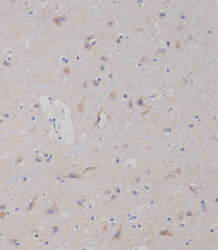
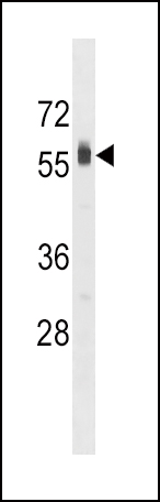
SPHK2 Antibody (C-term)
Purified Rabbit Polyclonal Antibody (Pab)
- 产品详情
- 文献引用 : 3
- 实验流程
- 背景知识
Application
| IHC-P-Leica, WB, E |
|---|---|
| Primary Accession | Q9NRA0 |
| Reactivity | Human, Mouse |
| Host | Rabbit |
| Clonality | Polyclonal |
| Isotype | Rabbit IgG |
| Calculated MW | 69217 Da |
| Antigen Region | 590-620 aa |
| Gene ID | 56848 |
|---|---|
| Other Names | Sphingosine kinase 2, SK 2, SPK 2, SPHK2 |
| Target/Specificity | This SPHK2 antibody is generated from rabbits immunized with a KLH conjugated synthetic peptide between 590-620 amino acids from the C-terminal region of human SPHK2. |
| Dilution | IHC-P-Leica~~1:250 WB~~1:1000 E~~Use at an assay dependent concentration. |
| Format | Purified polyclonal antibody supplied in PBS with 0.09% (W/V) sodium azide. This antibody is prepared by Saturated Ammonium Sulfate (SAS) precipitation followed by dialysis against PBS. |
| Storage | Maintain refrigerated at 2-8°C for up to 2 weeks. For long term storage store at -20°C in small aliquots to prevent freeze-thaw cycles. |
| Precautions | SPHK2 Antibody (C-term) is for research use only and not for use in diagnostic or therapeutic procedures. |
| Name | SPHK2 (HGNC:18859) |
|---|---|
| Synonyms | SK2 |
| Function | Catalyzes the phosphorylation of sphingosine to form sphingosine-1-phosphate (SPP), a lipid mediator with both intra- and extracellular functions. Also acts on D-erythro-dihydrosphingosine, D- erythro-sphingosine and L-threo-dihydrosphingosine. Binds phosphoinositides (PubMed:12954646, PubMed:19168031). In contrast to prosurvival SPHK1, has a positive effect on intracellular ceramide levels, inhibits cells growth and enhances apoptosis (PubMed:16118219). In mitochondria, is important for cytochrome-c oxidase assembly and mitochondrial respiration. The SPP produced in mitochondria binds PHB2 and modulates the regulation via PHB2 of complex IV assembly and respiration (PubMed:20959514). In nucleus, plays a role in epigenetic regulation of gene expression. Interacts with HDAC1 and HDAC2 and, through SPP production, inhibits their enzymatic activity, preventing the removal of acetyl groups from lysine residues with histones. Up- regulates acetylation of histone H3-K9, histone H4-K5 and histone H2B- K12 (PubMed:19729656). In nucleus, may have an inhibitory effect on DNA synthesis and cell cycle (PubMed:12954646, PubMed:16103110). In mast cells, is the main regulator of SPP production which mediates calcium influx, NF-kappa-B activation, cytokine production, such as TNF and IL6, and degranulation of mast cells (By similarity). In dopaminergic neurons, is involved in promoting mitochondrial functions regulating ATP and ROS levels (By similarity). Also involved in the regulation of glucose and lipid metabolism (By similarity). |
| Cellular Location | Cytoplasm. Nucleus. Endoplasmic reticulum {ECO:0000250|UniProtKB:Q9JIA7}. Mitochondrion inner membrane {ECO:0000250|UniProtKB:Q9JIA7}. Note=In nucleus, located in nucleosomes where it associates with core histone proteins such as histone 3 (PubMed:19729656). In brains of patients with Alzheimer's disease, may be preferentially localized in the nucleus. Cytosolic expression decrease correlates with the density of amyloid deposits (PubMed:29615132). In apoptotic cells, colocalizes with CASP1 in cell membrane where is cleaved and released from cells in an active form (PubMed:20197547). |
| Tissue Location | Mainly expressed in adult kidney, liver, and brain (PubMed:10751414). Expressed in cerebral cortex and hippocampus (at protein level) (PubMed:29615132). Isoform 1 is the predominant form expressed in most tissues (PubMed:16103110) |
For Research Use Only. Not For Use In Diagnostic Procedures.

Provided below are standard protocols that you may find useful for product applications.
BACKGROUND
Sphingosine Kinase (SphK) catalyzes the phosphorylation of the lipid sphingosine, creating the bioactive lipid sphingosine-1-phosphate (S1P). S1P subsequently signals through cell surface G protein-coupled receptors, as well as intracellularly, to modulate cell proliferation, survival, motility and differentiation. SphK is an important signaling enzyme which is activated by diverse agents, including growth factors that signal through receptor tyrosine kinases, agents activating G protein-coupled receptors, and immunoglobulin receptors. Two SphK isotypes, SphK-1 and SphK-2, have been cloned, and both isotypes are ubiquitously expressed. SphK-1 has been shown to mediate cell growth, prevention of apoptosis, and cellular transformation, and is upregulated in a variety of human tumors. In contrast, SphK-2 increases apoptosis, and may be responsible for phosphorylating and activating the immunosuppressive drug FTY720.
REFERENCES
Shu, X., et al., Mol. Cell. Biol. 22(22):7758-7768 (2002).
Xia, P., et al., J. Biol. Chem. 277(10):7996-8003 (2002).
Liu, H., et al., J. Biol. Chem. 275(26):19513-19520 (2000).
Shutler, G., et al., Genomics 34(3):334-339 (1996).
终于等到您。ABCEPTA(百远生物)抗体产品。
点击下方“我要评价 ”按钮提交您的反馈信息,您的反馈和评价是我们最宝贵的财富之一,
我们将在1-3个工作日内处理您的反馈信息。
如有疑问,联系:0512-88856768 tech-china@abcepta.com.


