


 癌症的基本特征包括细胞增殖、血管生成、迁移、凋亡逃避机制和细胞永生等。找到癌症发生过程中这些通路的关键标记物和对应的抗体用于检测至关重要。
 为您推荐一个泛素化位点预测神器——泛素化分析工具,可以为您的蛋白的泛素化位点作出预测和评分。
 细胞自噬受体图形绘图工具为你的蛋白的细胞受体结合位点作出预测和评分,识别结合到自噬通路中的蛋白是非常重要的,便于让我们理解自噬在正常生理、病理过程中的作用,如发育、细胞分化、神经退化性疾病、压力条件下、感染和癌症。
MSN Antibody
Purified Mouse Monoclonal Antibody
- 产品详情
- 实验流程
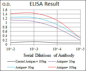
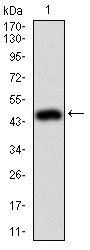
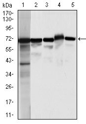
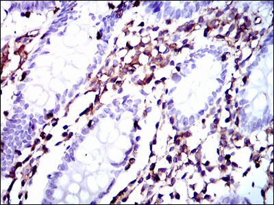
Application
| WB, IHC, FC, E |
|---|---|
| Primary Accession | P26038 |
| Reactivity | Human, Monkey |
| Host | Mouse |
| Clonality | Monoclonal |
| Clone Names | 2C12 |
| Isotype | IgG1 |
| Calculated MW | 67820 Da |
| Description | Moesin (for membrane-organizing extension spike protein) is a member of the ERM family which includes ezrin and radixin. ERM proteins appear to function as cross-linkers between plasma membranes and actin-based cytoskeletons. Moesin is localized to filopodia and other membranous protrusions that are important for cell-cell recognition and signaling and for cell movement. |
| Immunogen | Purified recombinant fragment of human MSN expressed in E. Coli. |
| Formulation | Purified antibody in PBS with 0.05% sodium azide |
| Gene ID | 4478 |
|---|---|
| Other Names | Moesin, Membrane-organizing extension spike protein, MSN |
| Dilution | WB~~1/500 - 1/2000 IHC~~1/200 - 1/1000 FC~~1/200 - 1/400 E~~1/10000 |
| Storage | Maintain refrigerated at 2-8°C for up to 6 months. For long term storage store at -20°C in small aliquots to prevent freeze-thaw cycles. |
| Precautions | MSN Antibody is for research use only and not for use in diagnostic or therapeutic procedures. |
| Name | MSN (HGNC:7373) |
|---|---|
| Function | Ezrin-radixin-moesin (ERM) family protein that connects the actin cytoskeleton to the plasma membrane and thereby regulates the structure and function of specific domains of the cell cortex. Tethers actin filaments by oscillating between a resting and an activated state providing transient interactions between moesin and the actin cytoskeleton (PubMed:10212266). Once phosphorylated on its C-terminal threonine, moesin is activated leading to interaction with F-actin and cytoskeletal rearrangement (PubMed:10212266). These rearrangements regulate many cellular processes, including cell shape determination, membrane transport, and signal transduction (PubMed:12387735, PubMed:15039356). The role of moesin is particularly important in immunity acting on both T and B-cells homeostasis and self-tolerance, regulating lymphocyte egress from lymphoid organs (PubMed:9298994, PubMed:9616160). Modulates phagolysosomal biogenesis in macrophages (By similarity). Also participates in immunologic synapse formation (PubMed:27405666). |
| Cellular Location | Cell membrane; Peripheral membrane protein {ECO:0000250|UniProtKB:P26041}; Cytoplasmic side {ECO:0000250|UniProtKB:P26041}. Cytoplasm, cytoskeleton {ECO:0000250|UniProtKB:P26041}. Apical cell membrane {ECO:0000250|UniProtKB:P26041}; Peripheral membrane protein {ECO:0000250|UniProtKB:P26041}; Cytoplasmic side {ECO:0000250|UniProtKB:P26041}. Cell projection, microvillus membrane {ECO:0000250|UniProtKB:P26041}; Peripheral membrane protein {ECO:0000250|UniProtKB:P26041}; Cytoplasmic side {ECO:0000250|UniProtKB:P26041}. Cell projection, microvillus {ECO:0000250|UniProtKB:P26041}. Note=Phosphorylated form is enriched in microvilli-like structures at apical membrane. Increased cell membrane localization of both phosphorylated and non-phosphorylated forms seen after thrombin treatment (By similarity). Localizes at the uropods of T lymphoblasts. {ECO:0000250|UniProtKB:P26041, ECO:0000269|PubMed:18586956, ECO:0000269|PubMed:9298994} |
| Tissue Location | In all tissues and cultured cells studied. |
Research Areas
For Research Use Only. Not For Use In Diagnostic Procedures.
Application Protocols
Provided below are standard protocols that you may find useful for product applications.
REFERENCES
Int J Cancer. 2009 Apr 1;124(7):1614-21. J Biol Chem. 2009 Jan 23;284(4):2419-34.
终于等到您。ABCEPTA(百远生物)抗体产品。
点击下方“我要评价 ”按钮提交您的反馈信息,您的反馈和评价是我们最宝贵的财富之一,
我们将在1-3个工作日内处理您的反馈信息。
如有疑问,联系:0512-88856768 tech-china@abcepta.com.
¥ 1,500.00
Cat# AO1688a


