


 癌症的基本特征包括细胞增殖、血管生成、迁移、凋亡逃避机制和细胞永生等。找到癌症发生过程中这些通路的关键标记物和对应的抗体用于检测至关重要。
 为您推荐一个泛素化位点预测神器——泛素化分析工具,可以为您的蛋白的泛素化位点作出预测和评分。
 细胞自噬受体图形绘图工具为你的蛋白的细胞受体结合位点作出预测和评分,识别结合到自噬通路中的蛋白是非常重要的,便于让我们理解自噬在正常生理、病理过程中的作用,如发育、细胞分化、神经退化性疾病、压力条件下、感染和癌症。
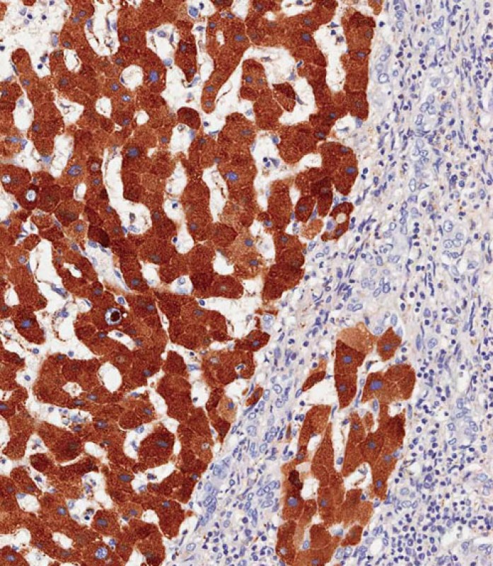
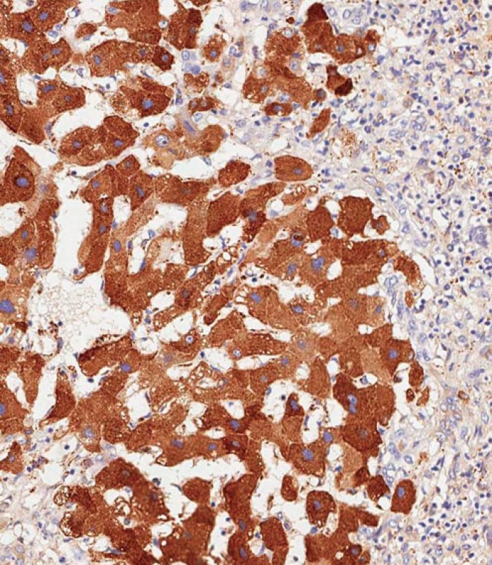
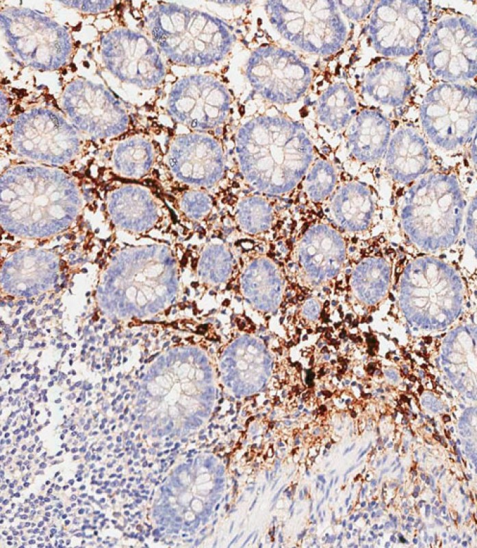
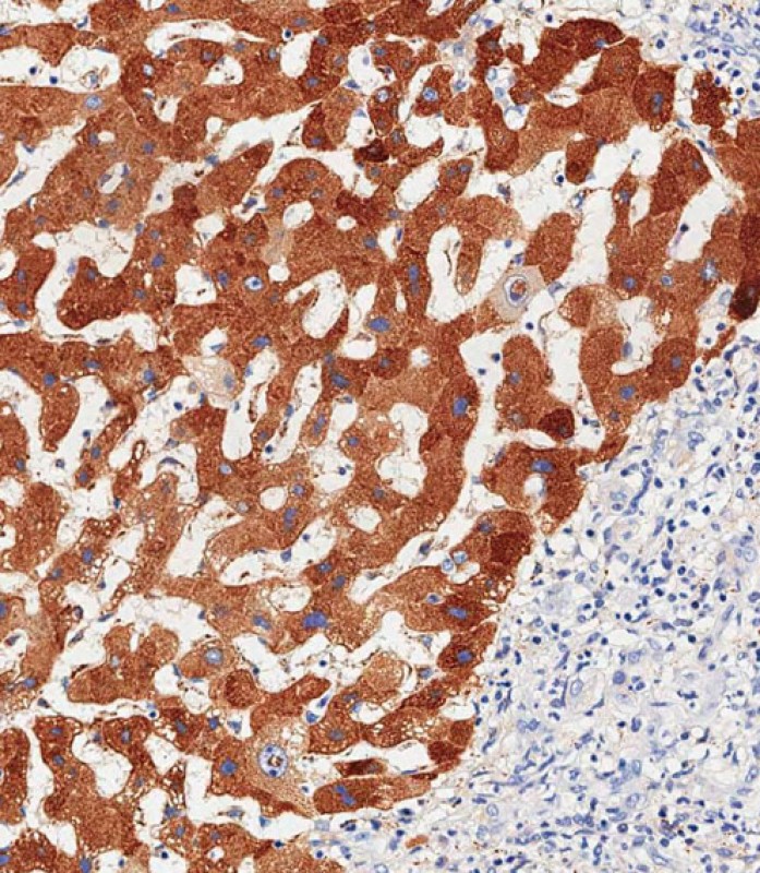
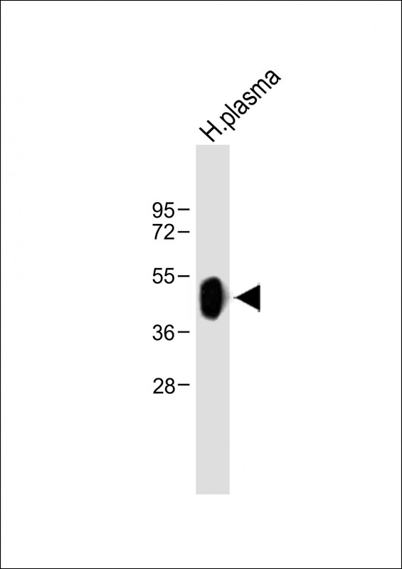
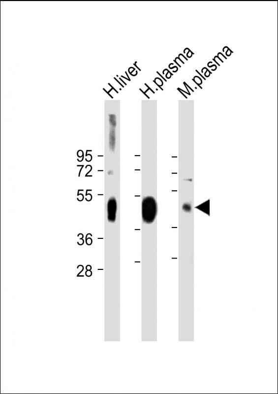
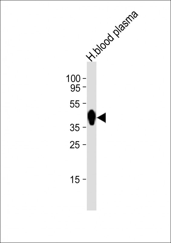
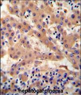
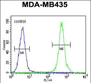
HP Antibody (Center)
Affinity Purified Rabbit Polyclonal Antibody (Pab)


- 产品详情
- 实验流程
- 背景知识
Application
| IHC-P-Leica, FC, IF, WB, E |
|---|---|
| Primary Accession | P00738 |
| Other Accession | NP_005134.1 |
| Reactivity | Human, Rat, Mouse |
| Host | Rabbit |
| Clonality | Polyclonal |
| Isotype | Rabbit IgG |
| Calculated MW | 45205 Da |
| Antigen Region | 296-322 aa |
| Gene ID | 3240 |
|---|---|
| Other Names | Haptoglobin, Zonulin, Haptoglobin alpha chain, Haptoglobin beta chain, HP |
| Target/Specificity | This HP antibody is generated from rabbits immunized with a KLH conjugated synthetic peptide between 296-322 amino acids from the Central region of human HP. |
| Dilution | IHC-P-Leica~~1:500 FC~~1:10~50 IF~~1:10~50 WB~~1:1000 E~~Use at an assay dependent concentration. |
| Format | Purified polyclonal antibody supplied in PBS with 0.09% (W/V) sodium azide. This antibody is purified through a protein A column, followed by peptide affinity purification. |
| Storage | Maintain refrigerated at 2-8°C for up to 2 weeks. For long term storage store at -20°C in small aliquots to prevent freeze-thaw cycles. |
| Precautions | HP Antibody (Center) is for research use only and not for use in diagnostic or therapeutic procedures. |
| Name | HP |
|---|---|
| Function | As a result of hemolysis, hemoglobin is found to accumulate in the kidney and is secreted in the urine. Haptoglobin captures, and combines with free plasma hemoglobin to allow hepatic recycling of heme iron and to prevent kidney damage. Haptoglobin also acts as an antioxidant, has antibacterial activity, and plays a role in modulating many aspects of the acute phase response. Hemoglobin/haptoglobin complexes are rapidly cleared by the macrophage CD163 scavenger receptor expressed on the surface of liver Kupfer cells through an endocytic lysosomal degradation pathway. |
| Cellular Location | Secreted. |
| Tissue Location | Expressed by the liver and secreted in plasma. |
For Research Use Only. Not For Use In Diagnostic Procedures.
Provided below are standard protocols that you may find useful for product applications.
BACKGROUND
This gene encodes a preproprotein, which is processed to yield both alpha and beta chains, which subsequently combine as a tetramer to produce haptoglobin. Haptoglobin functions to bind free plasma hemoglobin, which allows degradative enzymes to gain access to the hemoglobin, while at the same time preventing loss of iron through the kidneys and protecting the kidneys from damage by hemoglobin. Mutations in this gene and/or its regulatory regions cause ahaptoglobinemia or hypohaptoglobinemia. This gene has also been linked to diabetic nephropathy, the incidence of coronary artery disease in type 1 diabetes, Crohn's disease, inflammatory disease behavior, primary sclerosing cholangitis, susceptibility to idiopathic Parkinson's disease, and a reduced incidence of Plasmodium falciparum malaria. A similar duplicated gene is located next to this gene on chromosome 16. Multiple transcript variants encoding different isoforms have been found for this gene.
REFERENCES
Bailey, S.D., et al. Diabetes Care 33(10):2250-2253(2010)
Ucisik-Akkaya, E., et al. Mol. Hum. Reprod. 16(10):770-777(2010)
Ruano, G., et al. Pharmacogenomics 11(7):959-971(2010)
Savy, M., et al. PLoS ONE 5 (6), E11075 (2010) :
Kasvosve, I., et al. Adv Clin Chem 50, 23-46 (2010) :
终于等到您。ABCEPTA(百远生物)抗体产品。
点击下方“我要评价 ”按钮提交您的反馈信息,您的反馈和评价是我们最宝贵的财富之一,
我们将在1-3个工作日内处理您的反馈信息。
如有疑问,联系:0512-88856768 tech-china@abcepta.com.


