


 癌症的基本特征包括细胞增殖、血管生成、迁移、凋亡逃避机制和细胞永生等。找到癌症发生过程中这些通路的关键标记物和对应的抗体用于检测至关重要。
 为您推荐一个泛素化位点预测神器——泛素化分析工具,可以为您的蛋白的泛素化位点作出预测和评分。
 细胞自噬受体图形绘图工具为你的蛋白的细胞受体结合位点作出预测和评分,识别结合到自噬通路中的蛋白是非常重要的,便于让我们理解自噬在正常生理、病理过程中的作用,如发育、细胞分化、神经退化性疾病、压力条件下、感染和癌症。
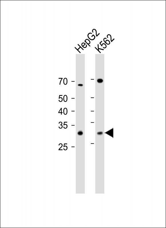
CLEC12B Antibody (C-term)
Affinity Purified Rabbit Polyclonal Antibody (Pab)

- 产品详情
- 实验流程
- 背景知识
Application
| WB, IHC-P, E |
|---|---|
| Primary Accession | Q2HXU8 |
| Other Accession | NP_001123470.1, NP_995324.2 |
| Reactivity | Human |
| Host | Rabbit |
| Clonality | Polyclonal |
| Isotype | Rabbit IgG |
| Calculated MW | 31616 Da |
| Antigen Region | 249-276 aa |
| Gene ID | 387837 |
|---|---|
| Other Names | C-type lectin domain family 12 member B, Macrophage antigen H, CLEC12B |
| Target/Specificity | This CLEC12B antibody is generated from rabbits immunized with a KLH conjugated synthetic peptide between 249-276 amino acids from the C-terminal region of human CLEC12B. |
| Dilution | WB~~1:1000 IHC-P~~1:100~500 E~~Use at an assay dependent concentration. |
| Format | Purified polyclonal antibody supplied in PBS with 0.05% (V/V) Proclin 300. This antibody is purified through a protein A column, followed by peptide affinity purification. |
| Storage | Maintain refrigerated at 2-8°C for up to 2 weeks. For long term storage store at -20°C in small aliquots to prevent freeze-thaw cycles. |
| Precautions | CLEC12B Antibody (C-term) is for research use only and not for use in diagnostic or therapeutic procedures. |
| Name | CLEC12B {ECO:0000303|PubMed:34310951, ECO:0000312|HGNC:HGNC:31966} |
|---|---|
| Function | Inhibitory receptor postulated to negatively regulate immune and non-immune functions (PubMed:17562706, PubMed:34310951). Upon phosphorylation, recruits SH2 domain-containing PTPN6 and PTPN11 phosphatases to its ITIM motif and antagonizes activation signals (PubMed:17562706, PubMed:34310951). Although it inhibits KLRK1/NKG2D- mediated signaling, it does not bind known ligands of KLRK1/NKG2D and therefore is not its inhibitory counterpart (PubMed:17562706). May limit activation of myeloid cell subsets in response to infection or tissue inflammation (PubMed:17562706). May protect target cells against natural killer cell-mediated lysis (PubMed:17562706). May negatively regulate cell cycle and differentiation of melanocytes via inactivation of STAT3 (PubMed:34310951). |
| Cellular Location | Cell membrane; Single-pass type II membrane protein |
| Tissue Location | Detected in colon, heart, kidney, liver, lung, mammary gland, ovary, spleen and testis (PubMed:17562706). Expressed in melanocytes (at protein level) (PubMed:34310951) |
Research Areas
For Research Use Only. Not For Use In Diagnostic Procedures.
Application Protocols
Provided below are standard protocols that you may find useful for product applications.
BACKGROUND
Cell surface receptor that protects target cells against natural killer cell-mediated lysis. Modulates signaling cascades and mediates tyrosine phosphorylation of target MAP kinases.
REFERENCES
Hoffmann, S.C., et al. J. Biol. Chem. 282(31):22370-22375(2007)
Clark, H.F., et al. Genome Res. 13(10):2265-2270(2003)
终于等到您。ABCEPTA(百远生物)抗体产品。
点击下方“我要评价 ”按钮提交您的反馈信息,您的反馈和评价是我们最宝贵的财富之一,
我们将在1-3个工作日内处理您的反馈信息。
如有疑问,联系:0512-88856768 tech-china@abcepta.com.
¥ 1,250.00
Cat# AP11585b


