


 癌症的基本特征包括细胞增殖、血管生成、迁移、凋亡逃避机制和细胞永生等。找到癌症发生过程中这些通路的关键标记物和对应的抗体用于检测至关重要。
 为您推荐一个泛素化位点预测神器——泛素化分析工具,可以为您的蛋白的泛素化位点作出预测和评分。
 细胞自噬受体图形绘图工具为你的蛋白的细胞受体结合位点作出预测和评分,识别结合到自噬通路中的蛋白是非常重要的,便于让我们理解自噬在正常生理、病理过程中的作用,如发育、细胞分化、神经退化性疾病、压力条件下、感染和癌症。
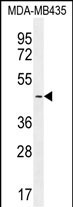
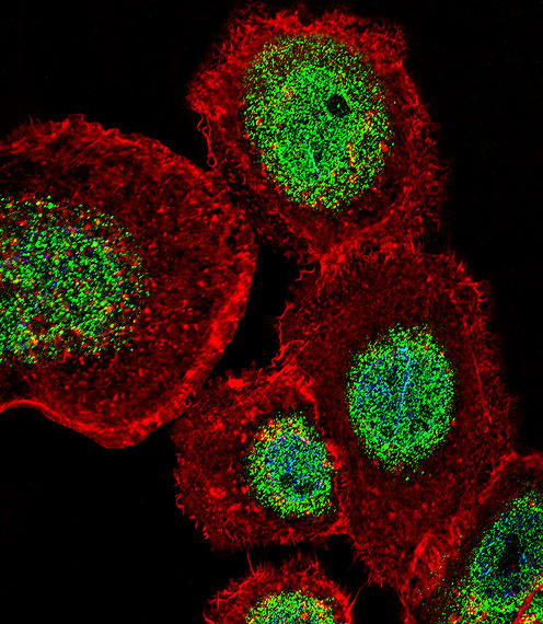
LEF1 Antibody (N-term)
Affinity Purified Rabbit Polyclonal Antibody (Pab)
- 产品详情
- 文献引用 : 3
- 实验流程
- 背景知识
Application
| WB, IF, E |
|---|---|
| Primary Accession | Q9UJU2 |
| Other Accession | Q9QXN1, P27782, NP_057353.1 |
| Reactivity | Human, Rat, Mouse |
| Predicted | Mouse, Rat |
| Host | Rabbit |
| Clonality | Polyclonal |
| Isotype | Rabbit IgG |
| Calculated MW | 44201 Da |
| Antigen Region | 10-37 aa |
| Gene ID | 51176 |
|---|---|
| Other Names | Lymphoid enhancer-binding factor 1, LEF-1, T cell-specific transcription factor 1-alpha, TCF1-alpha, LEF1 |
| Target/Specificity | This LEF1 antibody is generated from rabbits immunized with a KLH conjugated synthetic peptide between 10-37 amino acids from the N-terminal region of human LEF1. |
| Dilution | WB~~1:1000 IF~~1:10~50 E~~Use at an assay dependent concentration. |
| Format | Purified polyclonal antibody supplied in PBS with 0.09% (W/V) sodium azide. This antibody is purified through a protein A column, followed by peptide affinity purification. |
| Storage | Maintain refrigerated at 2-8°C for up to 2 weeks. For long term storage store at -20°C in small aliquots to prevent freeze-thaw cycles. |
| Precautions | LEF1 Antibody (N-term) is for research use only and not for use in diagnostic or therapeutic procedures. |
| Name | LEF1 (HGNC:6551) |
|---|---|
| Function | Transcription factor that binds DNA in a sequence-specific manner (PubMed:2010090). Participates in the Wnt signaling pathway (By similarity). Activates transcription of target genes in the presence of CTNNB1 and EP300 (By similarity). PIAG antagonizes both Wnt-dependent and Wnt-independent activation by LEF1 (By similarity). TLE1, TLE2, TLE3 and TLE4 repress transactivation mediated by LEF1 and CTNNB1 (PubMed:11266540). Regulates T-cell receptor alpha enhancer function (PubMed:19653274). Required for IL17A expressing gamma-delta T-cell maturation and development, via binding to regulator loci of BLK to modulate expression (By similarity). Acts as a positive regulator of odontoblast differentiation during mesenchymal tooth germ formation, expression is repressed during the bell stage by MSX1-mediated inhibition of CTNNB1 signaling (By similarity). May play a role in hair cell differentiation and follicle morphogenesis (By similarity). |
| Cellular Location | Nucleus {ECO:0000255|PROSITE-ProRule:PRU00267}. Note=Found in nuclear bodies upon PIASG binding. |
| Tissue Location | Detected in thymus. Not detected in normal colon, but highly expressed in colon cancer biopsies and colon cancer cell lines. Expressed in several pancreatic tumors and weakly expressed in normal pancreatic tissue. Isoforms 1 and 5 are detected in several pancreatic cell lines. |
For Research Use Only. Not For Use In Diagnostic Procedures.

Provided below are standard protocols that you may find useful for product applications.
BACKGROUND
This gene encodes a transcription factor belonging to a family of proteins that share homology with the high mobility group protein-1. The protein encoded by this gene can bind to a functionally important site in the T-cell receptor-alpha enhancer, thereby conferring maximal enhancer activity. This transcription factor is involved in the Wnt signaling pathway, and it may function in hair cell differentiation and follicle morphogenesis. Mutations in this gene have been found in somatic sebaceous tumors. This gene has also been linked to other cancers, including androgen-independent prostate cancer. Alternative splicing results in multiple transcript variants.
REFERENCES
Gutierrez, A. Jr., et al. Blood 116(16):2975-2983(2010)
Kalsi, G., et al. Hum. Mol. Genet. 19(12):2497-2506(2010)
Chen, Q.Y., et al. J. Immunol. 184(9):5047-5054(2010)
Beagle, B., et al. PLoS ONE 5 (7), E11821 (2010) :
Jugessur, A., et al. PLoS ONE 5 (7), E11493 (2010) :
终于等到您。ABCEPTA(百远生物)抗体产品。
点击下方“我要评价 ”按钮提交您的反馈信息,您的反馈和评价是我们最宝贵的财富之一,
我们将在1-3个工作日内处理您的反馈信息。
如有疑问,联系:0512-88856768 tech-china@abcepta.com.


